4HQJ

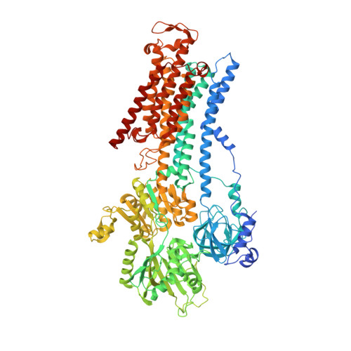
Crystal structure of Na+,K+-ATPase in the Na+-bound state


Experimental Data Snapshot

  • Method: X-RAY DIFFRACTION
  • Resolution: 4.30 Å
  • R-Value Free: 0.288 
  • R-Value Work: 0.261 
  • R-Value Observed: 0.263 

wwPDB Validation   3D Report Full Report


Ligand Structure Quality Assessment 


This is version 1.2 of the entry. See complete history


Literature

Crystal structure of Na+, K(+)-ATPase in the Na(+)-bound state.

Nyblom, M.Poulsen, H.Gourdon, P.Reinhard, L.Andersson, M.Lindahl, E.Fedosova, N.Nissen, P.

(2013) Science 342: 123-127

  • DOI: https://doi.org/10.1126/science.1243352
  • Primary Citation of Related Structures:  
    4HQJ

  • PubMed Abstract: 

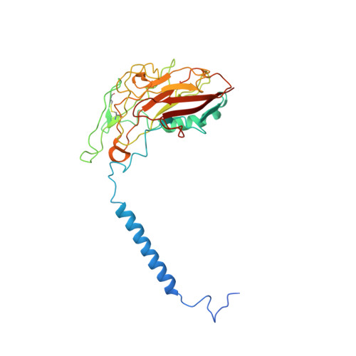
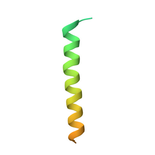
    The Na(+), K(+)-adenosine triphosphatase (ATPase) maintains the electrochemical gradients of Na(+) and K(+) across the plasma membrane--a prerequisite for electrical excitability and secondary transport. Hitherto, structural information has been limited to K(+)-bound or ouabain-blocked forms. We present the crystal structure of a Na(+)-bound Na(+), K(+)-ATPase as determined at 4.3 Å resolution. Compared with the K(+)-bound form, large conformational changes are observed in the α subunit whereas the β and γ subunit structures are maintained. The locations of the three Na(+) sites are indicated with the unique site III at the recently suggested IIIb, as further supported by electrophysiological studies on leak currents. Extracellular release of the third Na(+) from IIIb through IIIa, followed by exchange of Na(+) for K(+) at sites I and II, is suggested.


  • Organizational Affiliation

    Centre for Membrane Pumps in Cells and Disease-PUMPkin, Danish National Research Foundation, DK-8000 Aarhus, Denmark.


Macromolecules
Find similar proteins by:  (by identity cutoff)  |  3D Structure
Entity ID: 1
MoleculeChains Sequence LengthOrganismDetailsImage
Sodium/potassium-transporting ATPase subunit alpha-1A,
D [auth C]
1,021Sus scrofaMutation(s): 0 
EC: 3.6.3.9 (PDB Primary Data), 7.2.2.13 (UniProt)
Membrane Entity: Yes 
UniProt
Find proteins for P05024 (Sus scrofa)
Explore P05024 
Go to UniProtKB:  P05024
Entity Groups  
Sequence Clusters30% Identity50% Identity70% Identity90% Identity95% Identity100% Identity
UniProt GroupP05024
Sequence Annotations
Expand
  • Reference Sequence
Find similar proteins by:  (by identity cutoff)  |  3D Structure
Entity ID: 2
MoleculeChains Sequence LengthOrganismDetailsImage
Sodium/potassium-transporting ATPase subunit beta-1B,
E [auth D]
303Sus scrofaMutation(s): 0 
Membrane Entity: Yes 
UniProt
Find proteins for P05027 (Sus scrofa)
Explore P05027 
Go to UniProtKB:  P05027
Entity Groups  
Sequence Clusters30% Identity50% Identity70% Identity90% Identity95% Identity100% Identity
UniProt GroupP05027
Sequence Annotations
Expand
  • Reference Sequence
Find similar proteins by:  (by identity cutoff)  |  3D Structure
Entity ID: 3
MoleculeChains Sequence LengthOrganismDetailsImage
Na+/K+ ATPase gamma subunit transcript variant aC [auth G],
F [auth E]
65Sus scrofaMutation(s): 0 
Membrane Entity: Yes 
UniProt
Find proteins for Q58K79 (Sus scrofa)
Explore Q58K79 
Go to UniProtKB:  Q58K79
Entity Groups  
Sequence Clusters30% Identity50% Identity70% Identity90% Identity95% Identity100% Identity
UniProt GroupQ58K79
Sequence Annotations
Expand
  • Reference Sequence
Small Molecules
Ligands 5 Unique
IDChains Name / Formula / InChI Key2D Diagram3D Interactions
ADP
Query on ADP

Download Ideal Coordinates CCD File 
G [auth A],
O [auth C]
ADENOSINE-5'-DIPHOSPHATE
C10 H15 N5 O10 P2
XTWYTFMLZFPYCI-KQYNXXCUSA-N
CLR
Query on CLR

Download Ideal Coordinates CCD File 
M [auth A],
N [auth A],
U [auth C],
V [auth C],
W [auth D]
CHOLESTEROL
C27 H46 O
HVYWMOMLDIMFJA-DPAQBDIFSA-N
ALF
Query on ALF

Download Ideal Coordinates CCD File 
H [auth A],
P [auth C]
TETRAFLUOROALUMINATE ION
Al F4
UYOMQIYKOOHAMK-UHFFFAOYSA-J
MG
Query on MG

Download Ideal Coordinates CCD File 
I [auth A],
Q [auth C]
MAGNESIUM ION
Mg
JLVVSXFLKOJNIY-UHFFFAOYSA-N
NA
Query on NA

Download Ideal Coordinates CCD File 
J [auth A]
K [auth A]
L [auth A]
R [auth C]
S [auth C]
J [auth A],
K [auth A],
L [auth A],
R [auth C],
S [auth C],
T [auth C]
SODIUM ION
Na
FKNQFGJONOIPTF-UHFFFAOYSA-N
Experimental Data & Validation

Experimental Data

  • Method: X-RAY DIFFRACTION
  • Resolution: 4.30 Å
  • R-Value Free: 0.288 
  • R-Value Work: 0.261 
  • R-Value Observed: 0.263 
  • Space Group: P 21 21 21
Unit Cell:
Length ( Å )Angle ( ˚ )
a = 109.073α = 90
b = 219.578β = 90
c = 261.958γ = 90
Software Package:
Software NamePurpose
remdaq.pilatusdata collection
SHARPphasing
PHENIXrefinement
XDSdata reduction
XSCALEdata scaling

Structure Validation

View Full Validation Report


Ligand Structure Quality Assessment 


Entry History 

Deposition Data

Revision History  (Full details and data files)

  • Version 1.0: 2013-10-02
    Type: Initial release
  • Version 1.1: 2014-09-10
    Changes: Database references
  • Version 1.2: 2024-11-13
    Changes: Data collection, Database references, Derived calculations, Structure summary