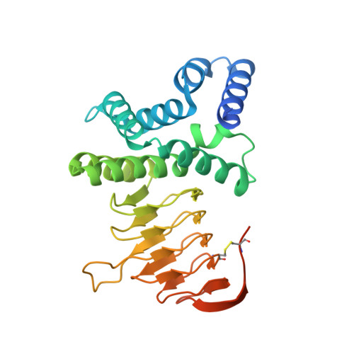
4HZC

Crystal structure of Serine acetyltransferase from Brucella abortus strain S19


Experimental Data Snapshot

  • Method: X-RAY DIFFRACTION
  • Resolution: 1.97 Å
  • R-Value Free: 0.239 
  • R-Value Work: 0.187 
  • R-Value Observed: 0.189 

wwPDB Validation   3D Report Full Report


This is version 1.4 of the entry. See complete history


Literature

Crystal structure of serine acetyl transferase from Brucella abortus and its complex with coenzyme A.

Kumar, S.Kumar, N.Alam, N.Gourinath, S.

(2014) Biochim Biophys Acta 1844: 1741-1748

  • DOI: https://doi.org/10.1016/j.bbapap.2014.07.009
  • Primary Citation of Related Structures:  
    4HZC, 4HZD

  • PubMed Abstract: 

    Brucella abortus is the major cause of premature foetal abortion in cattle, can be transmitted from cattle to humans, and is considered a powerful biological weapon. De novo cysteine biosynthesis is one of the essential pathways reported in bacteria, protozoa, and plants. Serine acetyltransferase (SAT) initiates this reaction by catalyzing the formation of O-acetylserine (OAS) using l-serine and acetyl coenzyme A as substrates. Here we report kinetic and crystallographic studies of this enzyme from B. abortus. The kinetic studies indicate that cysteine competitively inhibits the binding of serine to B. abortus SAT (BaSAT) and noncompetitively inhibits the binding of acetyl coenzyme A. The crystal structures of BaSAT in its apo state and in complex with coenzyme A (CoA) were determined to 1.96Å and 1.87Å resolution, respectively. BaSAT was observed as a trimer in a size exclusion column; however, it was seen as a hexamer in dynamic light scattering (DLS) studies and in the crystal structure, indicating it may exist in both states. The complex structure shows coenzyme A bound to the C-terminal region, making mostly hydrophobic contacts from the center of the active site extending up to the surface of the protein. There is no conformational difference in the enzyme between the apo and the complexed states, indicating lock and key binding and the absence of an induced fit mechanism.


  • Organizational Affiliation

    School of Life Sciences, Jawaharlal Nehru University, New Delhi, Delhi 110067, India.


Macromolecules
Find similar proteins by:  (by identity cutoff)  |  3D Structure
Entity ID: 1
MoleculeChains Sequence LengthOrganismDetailsImage
CysE, serine acetyltransferase
A, B, C, D, E
A, B, C, D, E, F, G, H, I, J, K, L
281Brucella abortus S19Mutation(s): 0 
Gene Names: BAbS19_I11940CysE
EC: 2.3.1.30
UniProt
Find proteins for A0A0F6AR69 (Brucella abortus (strain S19))
Explore A0A0F6AR69 
Go to UniProtKB:  A0A0F6AR69
Entity Groups  
Sequence Clusters30% Identity50% Identity70% Identity90% Identity95% Identity100% Identity
UniProt GroupA0A0F6AR69
Sequence Annotations
Expand
  • Reference Sequence
Small Molecules
Ligands 4 Unique
IDChains Name / Formula / InChI Key2D Diagram3D Interactions
15P
Query on 15P

Download Ideal Coordinates CCD File 
X [auth J]POLYETHYLENE GLYCOL (N=34)
C69 H140 O35
VUYXVWGKCKTUMF-UHFFFAOYSA-N
TRS
Query on TRS

Download Ideal Coordinates CCD File 
BA [auth K],
M [auth A],
Q [auth C],
S [auth D],
W [auth I]
2-AMINO-2-HYDROXYMETHYL-PROPANE-1,3-DIOL
C4 H12 N O3
LENZDBCJOHFCAS-UHFFFAOYSA-O
CL
Query on CL

Download Ideal Coordinates CCD File 
U [auth G]CHLORIDE ION
Cl
VEXZGXHMUGYJMC-UHFFFAOYSA-M
MG
Query on MG

Download Ideal Coordinates CCD File 
AA [auth K]
N [auth A]
O [auth A]
P [auth C]
R [auth D]
AA [auth K],
N [auth A],
O [auth A],
P [auth C],
R [auth D],
T [auth F],
V [auth G],
Y [auth J],
Z [auth J]
MAGNESIUM ION
Mg
JLVVSXFLKOJNIY-UHFFFAOYSA-N
Experimental Data & Validation

Experimental Data

  • Method: X-RAY DIFFRACTION
  • Resolution: 1.97 Å
  • R-Value Free: 0.239 
  • R-Value Work: 0.187 
  • R-Value Observed: 0.189 
  • Space Group: P 1 21 1
Unit Cell:
Length ( Å )Angle ( ˚ )
a = 73.697α = 90
b = 256.774β = 91.2
c = 82.285γ = 90
Software Package:
Software NamePurpose
DENZOdata reduction
SCALEPACKdata scaling
PHASERphasing
REFMACrefinement
PDB_EXTRACTdata extraction

Structure Validation

View Full Validation Report


Entry History 

Deposition Data

Revision History  (Full details and data files)

  • Version 1.0: 2014-02-19
    Type: Initial release
  • Version 1.1: 2014-08-06
    Changes: Database references
  • Version 1.2: 2014-08-13
    Changes: Database references
  • Version 1.3: 2017-11-15
    Changes: Refinement description
  • Version 1.4: 2024-10-30
    Changes: Data collection, Database references, Derived calculations, Structure summary