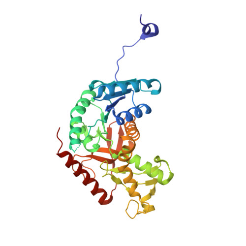
4I9H

Crystal structure of rabbit LDHA in complex with AP28669


Experimental Data Snapshot

  • Method: X-RAY DIFFRACTION
  • Resolution: 2.17 Å
  • R-Value Free: 0.243 
  • R-Value Work: 0.207 

Starting Model: experimental
View more details

wwPDB Validation   3D Report Full Report


Ligand Structure Quality Assessment 


This is version 1.2 of the entry. See complete history


Literature

Fragment growing and linking lead to novel nanomolar lactate dehydrogenase inhibitors.

Kohlmann, A.Zech, S.G.Li, F.Zhou, T.Squillace, R.M.Commodore, L.Greenfield, M.T.Lu, X.Miller, D.P.Huang, W.S.Qi, J.Thomas, R.M.Wang, Y.Zhang, S.Dodd, R.Liu, S.Xu, R.Xu, Y.Miret, J.J.Rivera, V.Clackson, T.Shakespeare, W.C.Zhu, X.Dalgarno, D.C.

(2013) J Med Chem 56: 1023-1040

  • DOI: https://doi.org/10.1021/jm3014844
  • Primary Citation of Related Structures:  
    4I8X, 4I9H, 4I9N, 4I9U

  • PubMed Abstract: 

    Lactate dehydrogenase A (LDH-A) catalyzes the interconversion of lactate and pyruvate in the glycolysis pathway. Cancer cells rely heavily on glycolysis instead of oxidative phosphorylation to generate ATP, a phenomenon known as the Warburg effect. The inhibition of LDH-A by small molecules is therefore of interest for potential cancer treatments. We describe the identification and optimization of LDH-A inhibitors by fragment-based drug discovery. We applied ligand based NMR screening to identify low affinity fragments binding to LDH-A. The dissociation constants (K(d)) and enzyme inhibition (IC(50)) of fragment hits were measured by surface plasmon resonance (SPR) and enzyme assays, respectively. The binding modes of selected fragments were investigated by X-ray crystallography. Fragment growing and linking, followed by chemical optimization, resulted in nanomolar LDH-A inhibitors that demonstrated stoichiometric binding to LDH-A. Selected molecules inhibited lactate production in cells, suggesting target-specific inhibition in cancer cell lines.


  • Organizational Affiliation

    ARIAD Pharmaceuticals, Inc., 26 Landsdowne Street, Cambridge, Massachusetts 02139, USA. anna.kohlmann@ariad.com


Macromolecules
Find similar proteins by:  (by identity cutoff)  |  3D Structure
Entity ID: 1
MoleculeChains Sequence LengthOrganismDetailsImage
L-lactate dehydrogenase A chain
A, B, C, D, E
A, B, C, D, E, F, G, H
331Oryctolagus cuniculusMutation(s): 0 
EC: 1.1.1.27
UniProt
Find proteins for P13491 (Oryctolagus cuniculus)
Explore P13491 
Go to UniProtKB:  P13491
Entity Groups  
Sequence Clusters30% Identity50% Identity70% Identity90% Identity95% Identity100% Identity
UniProt GroupP13491
Sequence Annotations
Expand
  • Reference Sequence
Small Molecules
Ligands 1 Unique
IDChains Name / Formula / InChI Key2D Diagram3D Interactions
1E4
Query on 1E4

Download Ideal Coordinates CCD File 
I [auth A]
J [auth B]
K [auth C]
L [auth D]
M [auth E]
I [auth A],
J [auth B],
K [auth C],
L [auth D],
M [auth E],
N [auth F],
O [auth G],
P [auth H]
1-O-[3-(5-carboxypyridin-2-yl)-5-fluorophenyl]-6-O-[4-({[(5-carboxypyridin-2-yl)sulfanyl]acetyl}amino)-2-chloro-5-methoxyphenyl]-D-mannitol
C33 H31 Cl F N3 O12 S
HTDBANWISVPXTR-KWINWIPXSA-N
Experimental Data & Validation

Experimental Data

  • Method: X-RAY DIFFRACTION
  • Resolution: 2.17 Å
  • R-Value Free: 0.243 
  • R-Value Work: 0.207 
  • Space Group: P 1 21 1
Unit Cell:
Length ( Å )Angle ( ˚ )
a = 83.866α = 90
b = 139.69β = 94.43
c = 138.81γ = 90
Software Package:
Software NamePurpose
CNSrefinement
PDB_EXTRACTdata extraction
StructureStudiodata collection
HKL-2000data reduction
HKL-2000data scaling
AMoREphasing

Structure Validation

View Full Validation Report


Ligand Structure Quality Assessment 


Entry History 

Deposition Data

Revision History  (Full details and data files)

  • Version 1.0: 2013-01-23
    Type: Initial release
  • Version 1.1: 2013-02-27
    Changes: Database references
  • Version 1.2: 2023-09-20
    Changes: Data collection, Database references, Derived calculations, Refinement description