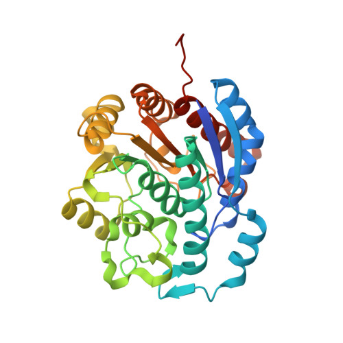
4IU4

Crystal structure of Leishmania mexicana arginase in complex with inhibitor BEC


Experimental Data Snapshot

  • Method: X-RAY DIFFRACTION
  • Resolution: 1.80 Å
  • R-Value Free: 0.193 
  • R-Value Work: 0.161 
  • R-Value Observed: 0.161 

Starting Model: experimental
View more details

wwPDB Validation   3D Report Full Report


Ligand Structure Quality Assessment 


This is version 1.3 of the entry. See complete history


Literature

Crystal structure of arginase from Leishmania mexicana and implications for the inhibition of polyamine biosynthesis in parasitic infections.

D'Antonio, E.L.Ullman, B.Roberts, S.C.Dixit, U.G.Wilson, M.E.Hai, Y.Christianson, D.W.

(2013) Arch Biochem Biophys 535: 163-176

  • DOI: https://doi.org/10.1016/j.abb.2013.03.015
  • Primary Citation of Related Structures:  
    4ITY, 4IU0, 4IU1, 4IU4, 4IU5

  • PubMed Abstract: 

    Arginase from parasitic protozoa belonging to the genus Leishmania is a potential drug target for the treatment of leishmaniasis because this binuclear manganese metalloenzyme catalyzes the first committed step in the biosynthesis of polyamines that enable cell growth and survival. The high resolution X-ray crystal structures of the unliganded form of Leishmania mexicana arginase (LmARG) and four inhibitor complexes are now reported. These complexes include the reactive substrate analogue 2(S)-amino-6-boronohexanoic acid (ABH) and the hydroxylated substrate analogue nor-N(ω)-hydroxy-l-arginine (nor-NOHA), which are the most potent arginase inhibitors known to date. Comparisons of the LmARG structure with that of the archetypal arginase, human arginase I, reveal that all residues important for substrate binding and catalysis are strictly conserved. However, three regions of tertiary structure differ between the parasitic enzyme and the human enzyme corresponding to the G62 - S71, L161 - C172, and I219 - V230 segments of LmARG. Additionally, variations are observed in salt link interactions that stabilize trimer assembly in LmARG. We also report biological studies in which we demonstrate that localization of LmARG to the glycosome, a unique subcellular organelle peculiar to Leishmania and related parasites, is essential for robust pathogenesis.


  • Organizational Affiliation

    Roy and Diana Vagelos Laboratories, Department of Chemistry, University of Pennsylvania, 231 South 34th Street, Philadelphia, PA 19104-6323, USA.


Macromolecules
Find similar proteins by:  (by identity cutoff)  |  3D Structure
Entity ID: 1
MoleculeChains Sequence LengthOrganismDetailsImage
Arginase330Leishmania mexicanaMutation(s): 0 
Gene Names: ARG
EC: 3.5.3.1
UniProt
Find proteins for Q6TUJ5 (Leishmania mexicana)
Explore Q6TUJ5 
Go to UniProtKB:  Q6TUJ5
Entity Groups  
Sequence Clusters30% Identity50% Identity70% Identity90% Identity95% Identity100% Identity
UniProt GroupQ6TUJ5
Sequence Annotations
Expand
  • Reference Sequence
Small Molecules
Ligands 4 Unique
IDChains Name / Formula / InChI Key2D Diagram3D Interactions
S2C
Query on S2C

Download Ideal Coordinates CCD File 
F [auth A]S-2-(BORONOETHYL)-L-CYSTEINE
C5 H13 B N O5 S
XLVRIIJULVQAMP-BYPYZUCNSA-N
GOL
Query on GOL

Download Ideal Coordinates CCD File 
D [auth A]GLYCEROL
C3 H8 O3
PEDCQBHIVMGVHV-UHFFFAOYSA-N
BME
Query on BME

Download Ideal Coordinates CCD File 
E [auth A]BETA-MERCAPTOETHANOL
C2 H6 O S
DGVVWUTYPXICAM-UHFFFAOYSA-N
MN
Query on MN

Download Ideal Coordinates CCD File 
B [auth A],
C [auth A]
MANGANESE (II) ION
Mn
WAEMQWOKJMHJLA-UHFFFAOYSA-N
Binding Affinity Annotations 
IDSourceBinding Affinity
S2C PDBBind:  4IU4 Ki: 1.00e+4 (nM) from 1 assay(s)
Experimental Data & Validation

Experimental Data

  • Method: X-RAY DIFFRACTION
  • Resolution: 1.80 Å
  • R-Value Free: 0.193 
  • R-Value Work: 0.161 
  • R-Value Observed: 0.161 
  • Space Group: H 3
Unit Cell:
Length ( Å )Angle ( ˚ )
a = 88.462α = 90
b = 88.462β = 90
c = 113.963γ = 120
Software Package:
Software NamePurpose
CBASSdata collection
CNSrefinement
HKL-2000data reduction
HKL-2000data scaling
CNSphasing

Structure Validation

View Full Validation Report


Ligand Structure Quality Assessment 


Entry History 

Deposition Data

Revision History  (Full details and data files)

  • Version 1.0: 2013-01-30
    Type: Initial release
  • Version 1.1: 2013-05-01
    Changes: Database references
  • Version 1.2: 2013-05-22
    Changes: Database references
  • Version 1.3: 2023-09-20
    Changes: Data collection, Database references, Derived calculations, Refinement description