4K4I

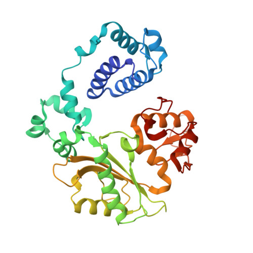
Ternary crystal structures of a human DNA POLYMERASE LAMBDA IN COMPLEX WITH DNA AND (-)FTC-TP.


Experimental Data Snapshot

  • Method: X-RAY DIFFRACTION
  • Resolution: 2.25 Å
  • R-Value Free: 0.274 
  • R-Value Work: 0.212 
  • R-Value Observed: 0.215 

wwPDB Validation   3D Report Full Report


Ligand Structure Quality Assessment 


This is version 1.3 of the entry. See complete history


Literature

Structural basis for the binding and incorporation of nucleotide analogs with L-stereochemistry by human DNA polymerase lambda.

Vyas, R.Zahurancik, W.J.Suo, Z.

(2014) Proc Natl Acad Sci U S A 111: E3033-E3042

  • DOI: https://doi.org/10.1073/pnas.1401286111
  • Primary Citation of Related Structures:  
    4K4G, 4K4H, 4K4I

  • PubMed Abstract: 

    Although lamivudine and emtricitabine, two L-deoxycytidine analogs, have been widely used as antiviral drugs for years, a structural basis for D-stereoselectivity against L-dNTPs, enantiomers of natural nucleotides (D-dNTPs), by any DNA polymerase or reverse transcriptase has not been established due to lack of a ternary structure of a polymerase, DNA, and an incoming L-dNTP. Here, we report 2.10-2.25 Å ternary crystal structures of human DNA polymerase λ, DNA, and L-deoxycytidine 5'-triphosphate (L-dCTP), or the triphosphates of lamivudine ((-)3TC-TP) and emtricitabine ((-)FTC-TP) with four ternary complexes per asymmetric unit. The structures of these 12 ternary complexes reveal that relative to D-deoxycytidine 5'-triphosphate (D-dCTP) in the canonical ternary structure of Polλ-DNA-D-dCTP, L-dCTP, (-)3TC-TP, and (-)FTC-TP all have their ribose rotated by 180°. Among the four ternary complexes with a specific L-nucleotide, two are similar and show that the L-nucleotide forms three Watson-Crick hydrogen bonds with the templating nucleotide dG and adopts a chair-like triphosphate conformation. In the remaining two similar ternary complexes, the L-nucleotide surprisingly interacts with the side chain of a conserved active site residue R517 through one or two hydrogen bonds, whereas the templating dG is anchored by a hydrogen bond with the side chain of a semiconserved residue Y505. Furthermore, the triphosphate of the L-nucleotide adopts an unprecedented N-shaped conformation. Our mutagenic and kinetic studies further demonstrate that the side chain of R517 is critical for the formation of the abovementioned four complexes along proposed catalytic pathways for L-nucleotide incorporation and provide the structural basis for the D-stereoselectivity of a DNA polymerase.


  • Organizational Affiliation

    Department of Chemistry and Biochemistry and.


Macromolecules

Find similar proteins by:  (by identity cutoff)  |  3D Structure
Entity ID: 1
MoleculeChains Sequence LengthOrganismDetailsImage
DNA polymerase lambda
A, E, I, M
340Homo sapiensMutation(s): 0 
Gene Names: POLL
EC: 2.7.7.7 (PDB Primary Data), 4.2.99 (PDB Primary Data)
UniProt & NIH Common Fund Data Resources
Find proteins for Q9UGP5 (Homo sapiens)
Explore Q9UGP5 
Go to UniProtKB:  Q9UGP5
PHAROS:  Q9UGP5
GTEx:  ENSG00000166169 
Entity Groups  
Sequence Clusters30% Identity50% Identity70% Identity90% Identity95% Identity100% Identity
UniProt GroupQ9UGP5
Sequence Annotations
Expand
  • Reference Sequence

Find similar nucleic acids by:  Sequence   |   3D Structure  

Entity ID: 2
MoleculeChains LengthOrganismImage
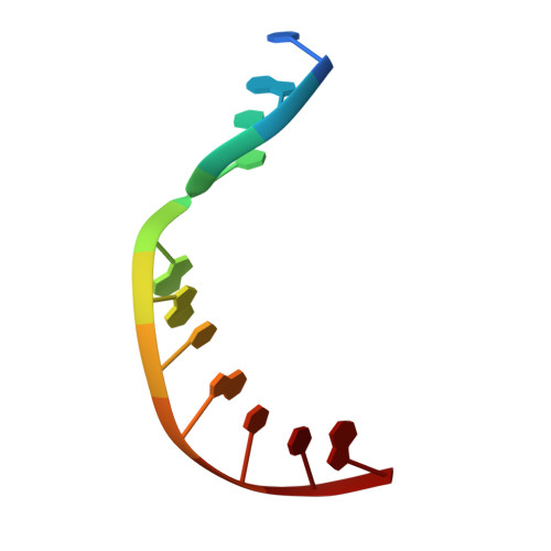
DNA (5'-D(*CP*GP*GP*CP*GP*GP*TP*AP*CP*TP*G)-3')
B, F, J, N
11N/A
Sequence Annotations
Expand
  • Reference Sequence

Find similar nucleic acids by:  Sequence   |   3D Structure  

Entity ID: 3
MoleculeChains LengthOrganismImage
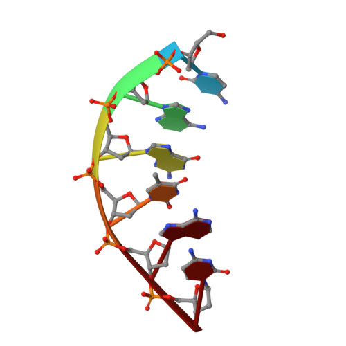
DNA (5'-D(*CP*AP*GP*TP*AP*C)-3')
C, G, K, O
6N/A
Sequence Annotations
Expand
  • Reference Sequence

Find similar nucleic acids by:  Sequence   |   3D Structure  

Entity ID: 4
MoleculeChains LengthOrganismImage
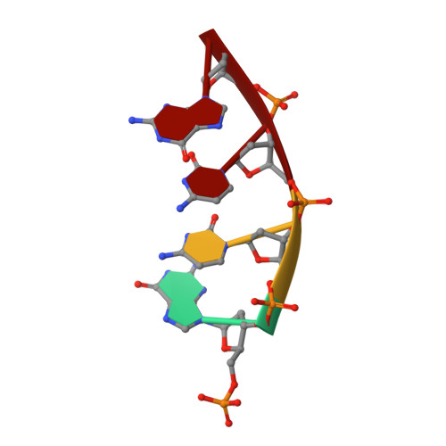
DNA (5'-D(P*GP*CP*CP*G)-3')
D, H, L, P
4N/A
Sequence Annotations
Expand
  • Reference Sequence
Small Molecules
Ligands 4 Unique
IDChains Name / Formula / InChI Key2D Diagram3D Interactions
1RY
Query on 1RY

Download Ideal Coordinates CCD File 
AA [auth E],
KA [auth I],
Q [auth A],
RA [auth M]
[[(2R,5S)-5-(4-azanyl-5-fluoranyl-2-oxidanylidene-pyrimidin-1-yl)-1,3-oxathiolan-2-yl]methoxy-oxidanyl-phosphoryl] phosphono hydrogen phosphate
C8 H13 F N3 O12 P3 S
WIEOLFZNMKSGEX-NTSWFWBYSA-N
CAC
Query on CAC

Download Ideal Coordinates CCD File 
GA [auth E],
WA [auth M]
CACODYLATE ION
C2 H6 As O2
OGGXGZAMXPVRFZ-UHFFFAOYSA-M
ACT
Query on ACT

Download Ideal Coordinates CCD File 
HA [auth E]
JA [auth F]
OA [auth I]
X [auth A]
XA [auth M]
HA [auth E],
JA [auth F],
OA [auth I],
X [auth A],
XA [auth M],
Z [auth B]
ACETATE ION
C2 H3 O2
QTBSBXVTEAMEQO-UHFFFAOYSA-M
CA
Query on CA

Download Ideal Coordinates CCD File 
BA [auth E]
CA [auth E]
DA [auth E]
EA [auth E]
FA [auth E]
BA [auth E],
CA [auth E],
DA [auth E],
EA [auth E],
FA [auth E],
IA [auth F],
LA [auth I],
MA [auth I],
NA [auth I],
PA [auth J],
QA [auth K],
R [auth A],
S [auth A],
SA [auth M],
T [auth A],
TA [auth M],
U [auth A],
UA [auth M],
V [auth A],
VA [auth M],
W [auth A],
Y [auth B],
YA [auth N]
CALCIUM ION
Ca
BHPQYMZQTOCNFJ-UHFFFAOYSA-N
Experimental Data & Validation

Experimental Data

  • Method: X-RAY DIFFRACTION
  • Resolution: 2.25 Å
  • R-Value Free: 0.274 
  • R-Value Work: 0.212 
  • R-Value Observed: 0.215 
  • Space Group: P 21 21 2
Unit Cell:
Length ( Å )Angle ( ˚ )
a = 194.44α = 90
b = 97.58β = 90
c = 105.15γ = 90
Software Package:
Software NamePurpose
PHASERphasing
REFMACrefinement
MOSFLMdata reduction
SCALAdata scaling

Structure Validation

View Full Validation Report


Ligand Structure Quality Assessment 


Entry History 

Deposition Data

Revision History  (Full details and data files)

  • Version 1.0: 2014-07-16
    Type: Initial release
  • Version 1.1: 2014-07-30
    Changes: Database references
  • Version 1.2: 2014-08-13
    Changes: Database references
  • Version 1.3: 2024-02-28
    Changes: Data collection, Database references, Derived calculations