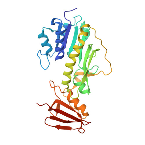
4LXU

dTdp-Fuc3N and 5-N-Formyl-THF


Experimental Data Snapshot

  • Method: X-RAY DIFFRACTION
  • Resolution: 1.40 Å
  • R-Value Free: 0.211 
  • R-Value Work: 0.180 
  • R-Value Observed: 0.182 

Starting Model: experimental
View more details

wwPDB Validation   3D Report Full Report


Ligand Structure Quality Assessment 


This is version 1.2 of the entry. See complete history


Literature

Structure of a sugar N-formyltransferase from Campylobacter jejuni.

Thoden, J.B.Goneau, M.F.Gilbert, M.Holden, H.M.

(2013) Biochemistry 52: 6114-6126

  • DOI: https://doi.org/10.1021/bi4009006
  • Primary Citation of Related Structures:  
    4LXQ, 4LXT, 4LXU, 4LXX, 4LXY, 4LY0, 4LY3

  • PubMed Abstract: 

    The O-antigens, which are components of the outer membranes of Gram-negative bacteria, are responsible for the wide species variations seen in nature and are thought to play a role in bacterial virulence. They often contain unusual dideoxysugars such as 3,6-dideoxy-3-formamido-d-glucose (Qui3NFo). Here, we describe a structural and functional investigation of the protein C8J_1081 from Campylobacter jejuni 81116, which is involved in the biosynthesis of Qui3NFo. Specifically, the enzyme, hereafter referred to as WlaRD, catalyzes the N-formylation of dTDP-3,6-dideoxy-3-amino-d-glucose (dTDP-Qui3N) using N(10)-formyltetrahydrofolate as the carbon source. For this investigation, seven X-ray structures of WlaRD, in complexes with various dTDP-linked sugars and cofactors, were determined to resolutions of 1.9 Å or better. One of the models, with bound N(10)-formyltetrahydrofolate and dTDP, represents the first glimpse of an N-formyltransferase with its natural cofactor. Another model contains the reaction products, tetrahydrofolate and dTDP-Qui3NFo. In combination, the structures provide snapshots of the WlaRD active site before and after catalysis. On the basis of these structures, three amino acid residues were targeted for study: Asn 94, His 96, and Asp 132. Mutations of any of these residues resulted in a complete loss of enzymatic activity. Given the position of His 96 in the active site, it can be postulated that it functions as the active site base to remove a proton from the sugar amino group as it attacks the carbonyl carbon of the N-10 formyl group of the cofactor. Enzyme assays demonstrate that WlaRD is also capable of utilizing dTDP-3,6-dideoxy-3-amino-d-galactose (dTDP-Fuc3N) as a substrate, albeit at a much reduced catalytic efficiency.


  • Organizational Affiliation

    Department of Biochemistry, University of Wisconsin , Madison, Wisconsin 53706, United States.


Macromolecules
Find similar proteins by:  (by identity cutoff)  |  3D Structure
Entity ID: 1
MoleculeChains Sequence LengthOrganismDetailsImage
WlaRD, a sugar 3N-formyl transferase
A, B
274Campylobacter jejuni subsp. jejuni 81116Mutation(s): 0 
Gene Names: C8J_1081
Sequence Annotations
Expand
  • Reference Sequence
Small Molecules
Ligands 6 Unique
IDChains Name / Formula / InChI Key2D Diagram3D Interactions
T3F
Query on T3F

Download Ideal Coordinates CCD File 
E [auth A],
P [auth B]
(3R,4S,5R,6R)-4-amino-3,5-dihydroxy-6-methyloxan-2-yl][hydroxy-[[(2R,3S,5R)-3-hydroxy-5-(5-methyl-2,4-dioxopyrimidin-1-yl)oxolan-2-yl]methoxy]phosphoryl] hydrogen phosphate
C16 H27 N3 O14 P2
KVYJLJOGNUNRJK-FQLHZTMTSA-N
FON
Query on FON

Download Ideal Coordinates CCD File 
C [auth A],
N [auth B]
N-{[4-({[(6R)-2-amino-5-formyl-4-oxo-1,4,5,6,7,8-hexahydropteridin-6-yl]methyl}amino)phenyl]carbonyl}-L-glutamic acid
C20 H23 N7 O7
VVIAGPKUTFNRDU-OLZOCXBDSA-N
MPO
Query on MPO

Download Ideal Coordinates CCD File 
D [auth A],
O [auth B]
3[N-MORPHOLINO]PROPANE SULFONIC ACID
C7 H15 N O4 S
DVLFYONBTKHTER-UHFFFAOYSA-N
SO4
Query on SO4

Download Ideal Coordinates CCD File 
M [auth A]SULFATE ION
O4 S
QAOWNCQODCNURD-UHFFFAOYSA-L
EDO
Query on EDO

Download Ideal Coordinates CCD File 
F [auth A]
G [auth A]
H [auth A]
I [auth A]
J [auth A]
F [auth A],
G [auth A],
H [auth A],
I [auth A],
J [auth A],
Q [auth B],
R [auth B],
S [auth B],
T [auth B],
U [auth B]
1,2-ETHANEDIOL
C2 H6 O2
LYCAIKOWRPUZTN-UHFFFAOYSA-N
CL
Query on CL

Download Ideal Coordinates CCD File 
K [auth A],
L [auth A]
CHLORIDE ION
Cl
VEXZGXHMUGYJMC-UHFFFAOYSA-M
Experimental Data & Validation

Experimental Data

  • Method: X-RAY DIFFRACTION
  • Resolution: 1.40 Å
  • R-Value Free: 0.211 
  • R-Value Work: 0.180 
  • R-Value Observed: 0.182 
  • Space Group: C 1 2 1
Unit Cell:
Length ( Å )Angle ( ˚ )
a = 96.534α = 90
b = 64.884β = 110.67
c = 134.458γ = 90
Software Package:
Software NamePurpose
HKL-3000data collection
PHASERphasing
REFMACrefinement
HKL-3000data reduction
HKL-3000data scaling

Structure Validation

View Full Validation Report


Ligand Structure Quality Assessment 


Entry History 

Deposition Data

Revision History  (Full details and data files)

  • Version 1.0: 2013-08-14
    Type: Initial release
  • Version 1.1: 2014-02-05
    Changes: Database references
  • Version 1.2: 2023-09-20
    Changes: Data collection, Database references, Derived calculations, Refinement description