4MUK

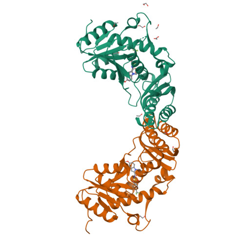
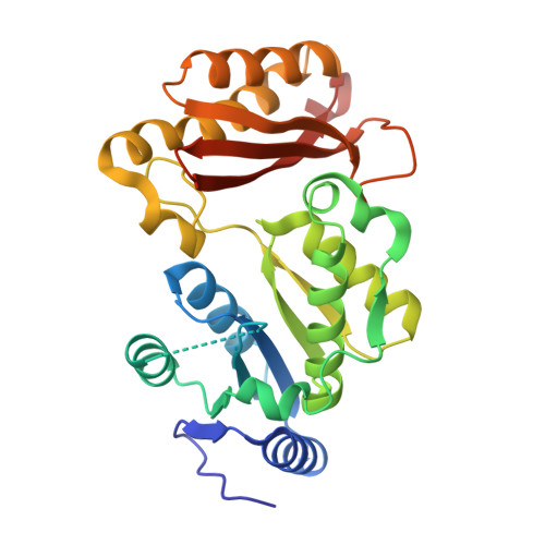
Crystal structure of pantothenate synthetase in complex with 2-(5-methoxy-2-(4-(trifluoromethyl)benzylsulfonylcarbamoyl)-1H-indol-1-yl)acetic acid


Experimental Data Snapshot

  • Method: X-RAY DIFFRACTION
  • Resolution: 1.90 Å
  • R-Value Free: 0.231 
  • R-Value Work: 0.174 
  • R-Value Observed: 0.177 

Starting Model: experimental
View more details

wwPDB Validation   3D Report Full Report


Ligand Structure Quality Assessment 


This is version 1.2 of the entry. See complete history


Literature

Optimization of Inhibitors of Mycobacterium tuberculosis Pantothenate Synthetase Based on Group Efficiency Analysis.

Hung, A.W.Silvestre, H.L.Wen, S.George, G.P.Boland, J.Blundell, T.L.Ciulli, A.Abell, C.

(2016) ChemMedChem 11: 38-42

  • DOI: https://doi.org/10.1002/cmdc.201500414
  • Primary Citation of Related Structures:  
    4MQ6, 4MUE, 4MUF, 4MUG, 4MUH, 4MUI, 4MUJ, 4MUK, 4MUL, 4MUN

  • PubMed Abstract: 

    Ligand efficiency has proven to be a valuable concept for optimization of leads in the early stages of drug design. Taking this one step further, group efficiency (GE) evaluates the binding efficiency of each appendage of a molecule, further fine-tuning the drug design process. Here, GE analysis is used to systematically improve the potency of inhibitors of Mycobacterium tuberculosis pantothenate synthetase, an important target in tuberculosis therapy. Binding efficiencies were found to be distributed unevenly within a lead molecule derived using a fragment-based approach. Substitution of the less efficient parts of the molecule allowed systematic development of more potent compounds. This method of dissecting and analyzing different groups within a molecule offers a rational and general way of carrying out lead optimization, with potential broad application within drug discovery.


  • Organizational Affiliation

    Department of Chemistry, University of Cambridge, Lensfield Rd, Cambridge, CB2 1EW, UK.


Macromolecules
Find similar proteins by:  (by identity cutoff)  |  3D Structure
Entity ID: 1
MoleculeChains Sequence LengthOrganismDetailsImage
Pantothenate synthetase
A, B
300Mycobacterium tuberculosisMutation(s): 2 
Gene Names: MT3707MTCY07H7B.20panCRv3602c
EC: 6.3.2.1
Sequence Annotations
Expand
  • Reference Sequence
Small Molecules
Ligands 3 Unique
IDChains Name / Formula / InChI Key2D Diagram3D Interactions
2DV
Query on 2DV

Download Ideal Coordinates CCD File 
C [auth A],
L [auth B]
[5-methoxy-2-({[4-(trifluoromethyl)benzyl]sulfonyl}carbamoyl)-1H-indol-1-yl]acetic acid
C20 H17 F3 N2 O6 S
BCONJIVPNWPJDO-UHFFFAOYSA-N
EDO
Query on EDO

Download Ideal Coordinates CCD File 
G [auth A],
H [auth A],
I [auth A],
J [auth A],
M [auth B]
1,2-ETHANEDIOL
C2 H6 O2
LYCAIKOWRPUZTN-UHFFFAOYSA-N
EOH
Query on EOH

Download Ideal Coordinates CCD File 
D [auth A],
E [auth A],
F [auth A],
K [auth A],
N [auth B]
ETHANOL
C2 H6 O
LFQSCWFLJHTTHZ-UHFFFAOYSA-N
Binding Affinity Annotations 
IDSourceBinding Affinity
2DV BindingDB:  4MUK IC50: 250 (nM) from 1 assay(s)
Experimental Data & Validation

Experimental Data

  • Method: X-RAY DIFFRACTION
  • Resolution: 1.90 Å
  • R-Value Free: 0.231 
  • R-Value Work: 0.174 
  • R-Value Observed: 0.177 
  • Space Group: P 1 21 1
Unit Cell:
Length ( Å )Angle ( ˚ )
a = 48.62α = 90
b = 70.91β = 99.51
c = 81.81γ = 90
Software Package:
Software NamePurpose
MAR345dtbdata collection
AMoREphasing
REFMACrefinement
MOSFLMdata reduction
SCALAdata scaling

Structure Validation

View Full Validation Report


Ligand Structure Quality Assessment 


Entry History 

Deposition Data

Revision History  (Full details and data files)

  • Version 1.0: 2014-08-27
    Type: Initial release
  • Version 1.1: 2016-11-23
    Changes: Database references
  • Version 1.2: 2023-09-20
    Changes: Data collection, Database references, Derived calculations, Refinement description