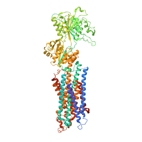
4NAB

Structure of the (SR)Ca2+-ATPase mutant E309Q in the Ca2-E1-MgAMPPCP form


Experimental Data Snapshot

  • Method: X-RAY DIFFRACTION
  • Resolution: 3.50 Å
  • R-Value Free: 0.266 
  • R-Value Work: 0.216 
  • R-Value Observed: 0.218 

wwPDB Validation   3D Report Full Report


Ligand Structure Quality Assessment 


This is version 1.3 of the entry. See complete history


Literature

SERCA mutant E309Q binds two Ca(2+) ions but adopts a catalytically incompetent conformation.

Clausen, J.D.Bublitz, M.Arnou, B.Montigny, C.Jaxel, C.Moller, J.V.Nissen, P.Andersen, J.P.le Maire, M.

(2013) EMBO J 32: 3231-3243

  • DOI: https://doi.org/10.1038/emboj.2013.250
  • Primary Citation of Related Structures:  
    4NAB

  • PubMed Abstract: 

    The sarco(endo)plasmic reticulum Ca(2+)-ATPase (SERCA) couples ATP hydrolysis to transport of Ca(2+). This directed energy transfer requires cross-talk between the two Ca(2+) sites and the phosphorylation site over 50 Å distance. We have addressed the mechano-structural basis for this intramolecular signal by analysing the structure and the functional properties of SERCA mutant E309Q. Glu(309) contributes to Ca(2+) coordination at site II, and a consensus has been that E309Q only binds Ca(2+) at site I. The crystal structure of E309Q in the presence of Ca(2+) and an ATP analogue, however, reveals two occupied Ca(2+) sites of a non-catalytic Ca2E1 state. Ca(2+) is bound with micromolar affinity by both Ca(2+) sites in E309Q, but without cooperativity. The Ca(2+)-bound mutant does phosphorylate from ATP, but at a very low maximal rate. Phosphorylation depends on the correct positioning of the A-domain, requiring a shift of transmembrane segment M1 into an 'up and kinked position'. This transition is impaired in the E309Q mutant, most likely due to a lack of charge neutralization and altered hydrogen binding capacities at Ca(2+) site II.


  • Organizational Affiliation

    1] Department of Molecular Biology and Genetics, Centre for Membrane Pumps in Cells and Disease - PUMPKIN, Danish National Research Foundation, Aarhus University, Aarhus, Denmark [2] Department of Biomedicine, Aarhus University, Aarhus, Denmark.


Macromolecules
Find similar proteins by:  (by identity cutoff)  |  3D Structure
Entity ID: 1
MoleculeChains Sequence LengthOrganismDetailsImage
Sarcoplasmic/endoplasmic reticulum calcium ATPase 11,000Oryctolagus cuniculusMutation(s): 1 
Gene Names: ATP2A1
EC: 3.6.3.8 (PDB Primary Data), 7.2.2.10 (UniProt)
Membrane Entity: Yes 
UniProt
Find proteins for P04191 (Oryctolagus cuniculus)
Explore P04191 
Go to UniProtKB:  P04191
Entity Groups  
Sequence Clusters30% Identity50% Identity70% Identity90% Identity95% Identity100% Identity
UniProt GroupP04191
Sequence Annotations
Expand
  • Reference Sequence
Experimental Data & Validation

Experimental Data

  • Method: X-RAY DIFFRACTION
  • Resolution: 3.50 Å
  • R-Value Free: 0.266 
  • R-Value Work: 0.216 
  • R-Value Observed: 0.218 
  • Space Group: C 1 2 1
Unit Cell:
Length ( Å )Angle ( ˚ )
a = 167.01α = 90
b = 55.79β = 109.29
c = 161.79γ = 90
Software Package:
Software NamePurpose
remdaq.pilatusdata collection
PHASERphasing
PHENIXrefinement
XDSdata reduction
XSCALEdata scaling

Structure Validation

View Full Validation Report


Ligand Structure Quality Assessment 


Entry History 

Deposition Data

Revision History  (Full details and data files)

  • Version 1.0: 2013-12-18
    Type: Initial release
  • Version 1.1: 2014-02-05
    Changes: Database references
  • Version 1.2: 2017-08-09
    Changes: Database references, Structure summary
  • Version 1.3: 2024-11-20
    Changes: Data collection, Database references, Derived calculations, Structure summary