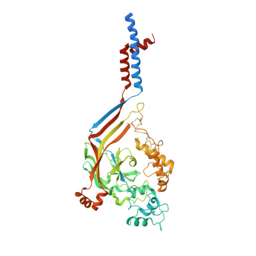
4NYK

Structure of a membrane protein


Experimental Data Snapshot

  • Method: X-RAY DIFFRACTION
  • Resolution: 3.00 Å
  • R-Value Free: 0.246 
  • R-Value Work: 0.202 
  • R-Value Observed: 0.204 

wwPDB Validation   3D Report Full Report


This is version 1.1 of the entry. See complete history


Literature

Pore architecture and ion sites in acid-sensing ion channels and P2X receptors.

Gonzales, E.B.Kawate, T.Gouaux, E.

(2009) Nature 460: 599-604

  • DOI: https://doi.org/10.1038/nature08218
  • Primary Citation of Related Structures:  
    3IJ4, 4NYK

  • PubMed Abstract: 

    Acid-sensing ion channels are proton-activated, sodium-selective channels composed of three subunits, and are members of the superfamily of epithelial sodium channels, mechanosensitive and FMRF-amide peptide-gated ion channels. These ubiquitous eukaryotic ion channels have essential roles in biological activities as diverse as sodium homeostasis, taste and pain. Despite their crucial roles in biology and their unusual trimeric subunit stoichiometry, there is little knowledge of the structural and chemical principles underlying their ion channel architecture and ion-binding sites. Here we present the structure of a functional acid-sensing ion channel in a desensitized state at 3 A resolution, the location and composition of the approximately 8 A 'thick' desensitization gate, and the trigonal antiprism coordination of caesium ions bound in the extracellular vestibule. Comparison of the acid-sensing ion channel structure with the ATP-gated P2X(4) receptor reveals similarity in pore architecture and aqueous vestibules, suggesting that there are unanticipated yet common structural and mechanistic principles.


  • Organizational Affiliation

    Vollum Institute, Oregon Health and Science University, 3181 Southwest Sam Jackson Park Road, Portland, Oregon 97239, USA.


Macromolecules
Find similar proteins by:  (by identity cutoff)  |  3D Structure
Entity ID: 1
MoleculeChains Sequence LengthOrganismDetailsImage
Acid-sensing ion channel 1470Gallus gallusMutation(s): 0 
Gene Names: ASIC1ACCN2
Membrane Entity: Yes 
UniProt
Find proteins for Q1XA76 (Gallus gallus)
Explore Q1XA76 
Go to UniProtKB:  Q1XA76
Entity Groups  
Sequence Clusters30% Identity50% Identity70% Identity90% Identity95% Identity100% Identity
UniProt GroupQ1XA76
Sequence Annotations
Expand
  • Reference Sequence
Small Molecules
Ligands 1 Unique
IDChains Name / Formula / InChI Key2D Diagram3D Interactions
CL
Query on CL

Download Ideal Coordinates CCD File 
B [auth A]CHLORIDE ION
Cl
VEXZGXHMUGYJMC-UHFFFAOYSA-M
Experimental Data & Validation

Experimental Data

  • Method: X-RAY DIFFRACTION
  • Resolution: 3.00 Å
  • R-Value Free: 0.246 
  • R-Value Work: 0.202 
  • R-Value Observed: 0.204 
  • Space Group: H 3
Unit Cell:
Length ( Å )Angle ( ˚ )
a = 131.787α = 90
b = 131.787β = 90
c = 119.107γ = 120
Software Package:
Software NamePurpose
ADSCdata collection
PHASERphasing
PHENIXrefinement
HKL-2000data reduction
HKL-2000data scaling

Structure Validation

View Full Validation Report


Entry History 

Deposition Data

Revision History  (Full details and data files)

  • Version 1.0: 2014-02-12
    Type: Initial release
  • Version 1.1: 2024-11-06
    Changes: Data collection, Database references, Derived calculations, Structure summary