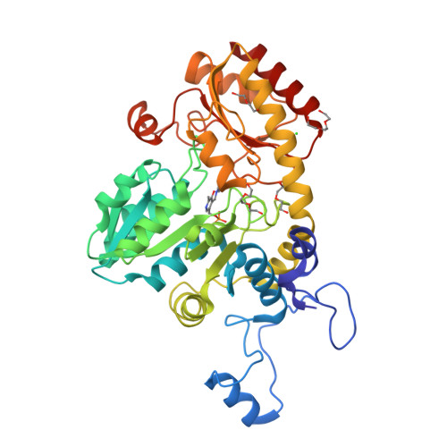
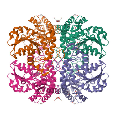
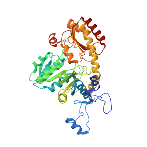
4OMA

The crystal structure of methionine gamma-lyase from Citrobacter freundii in complex with L-cycloserine pyridoxal-5'-phosphate


Experimental Data Snapshot

  • Method: X-RAY DIFFRACTION
  • Resolution: 1.60 Å
  • R-Value Free: 0.197 
  • R-Value Work: 0.139 
  • R-Value Observed: 0.142 

Starting Model: experimental
View more details

wwPDB Validation   3D Report Full Report


Ligand Structure Quality Assessment 


This is version 1.3 of the entry. See complete history


Literature

Pre-steady-state Kinetic and Structural Analysis of Interaction of Methionine gamma-Lyase from Citrobacter freundii with Inhibitors.

Kuznetsov, N.A.Faleev, N.G.Kuznetsova, A.A.Morozova, E.A.Revtovich, S.V.Anufrieva, N.V.Nikulin, A.D.Fedorova, O.S.Demidkina, T.V.

(2015) J Biol Chem 290: 671-681

  • DOI: https://doi.org/10.1074/jbc.M114.586511
  • Primary Citation of Related Structures:  
    4OMA

  • PubMed Abstract: 

    Methionine γ-lyase (MGL) catalyzes the γ-elimination of l-methionine and its derivatives as well as the β-elimination of l-cysteine and its analogs. These reactions yield α-keto acids and thiols. The mechanism of chemical conversion of amino acids includes numerous reaction intermediates. The detailed analysis of MGL interaction with glycine, l-alanine, l-norvaline, and l-cycloserine was performed by pre-steady-state stopped-flow kinetics. The structure of side chains of the amino acids is important both for their binding with enzyme and for the stability of the external aldimine and ketimine intermediates. X-ray structure of the MGL·l-cycloserine complex has been solved at 1.6 Å resolution. The structure models the ketimine intermediate of physiological reaction. The results elucidate the mechanisms of the intermediate interconversion at the stages of external aldimine and ketimine formation.


  • Organizational Affiliation

    From the Institute of Chemical Biology and Fundamental Medicine, Siberian Branch of the Russian Academy of Sciences, Novosibirsk 630090, the Department of Natural Sciences, Novosibirsk State University, Novosibirsk 630090.


Macromolecules
Find similar proteins by:  (by identity cutoff)  |  3D Structure
Entity ID: 1
MoleculeChains Sequence LengthOrganismDetailsImage
methionine gamma-lyase398Citrobacter freundiiMutation(s): 0 
Gene Names: H922_06139megl
EC: 4.4.1.11
UniProt
Find proteins for Q84AR1 (Citrobacter freundii)
Explore Q84AR1 
Go to UniProtKB:  Q84AR1
Entity Groups  
Sequence Clusters30% Identity50% Identity70% Identity90% Identity95% Identity100% Identity
UniProt GroupQ84AR1
Sequence Annotations
Expand
  • Reference Sequence
Small Molecules
Ligands 4 Unique
IDChains Name / Formula / InChI Key2D Diagram3D Interactions
LCS
Query on LCS

Download Ideal Coordinates CCD File 
B [auth A][5-hydroxy-6-methyl-4-({[(4E)-3-oxo-1,2-oxazolidin-4-ylidene]amino}methyl)pyridin-3-yl]methyl dihydrogen phosphate
C11 H14 N3 O7 P
KFCQHWOGBVCKHR-UKTHLTGXSA-N
PGE
Query on PGE

Download Ideal Coordinates CCD File 
C [auth A],
D [auth A]
TRIETHYLENE GLYCOL
C6 H14 O4
ZIBGPFATKBEMQZ-UHFFFAOYSA-N
PEG
Query on PEG

Download Ideal Coordinates CCD File 
E [auth A],
F [auth A]
DI(HYDROXYETHYL)ETHER
C4 H10 O3
MTHSVFCYNBDYFN-UHFFFAOYSA-N
CL
Query on CL

Download Ideal Coordinates CCD File 
G [auth A]CHLORIDE ION
Cl
VEXZGXHMUGYJMC-UHFFFAOYSA-M
Experimental Data & Validation

Experimental Data

  • Method: X-RAY DIFFRACTION
  • Resolution: 1.60 Å
  • R-Value Free: 0.197 
  • R-Value Work: 0.139 
  • R-Value Observed: 0.142 
  • Space Group: I 2 2 2
Unit Cell:
Length ( Å )Angle ( ˚ )
a = 56.27α = 90
b = 122.89β = 90
c = 126.61γ = 90
Software Package:
Software NamePurpose
MAR345dtbdata collection
PHASESphasing
REFMACrefinement
PROCESSdata reduction
PROCESSdata scaling

Structure Validation

View Full Validation Report


Ligand Structure Quality Assessment 


Entry History 

Deposition Data

Revision History  (Full details and data files)

  • Version 1.0: 2014-11-26
    Type: Initial release
  • Version 1.1: 2014-12-03
    Changes: Database references
  • Version 1.2: 2015-01-21
    Changes: Database references
  • Version 1.3: 2023-09-20
    Changes: Data collection, Database references, Derived calculations, Refinement description