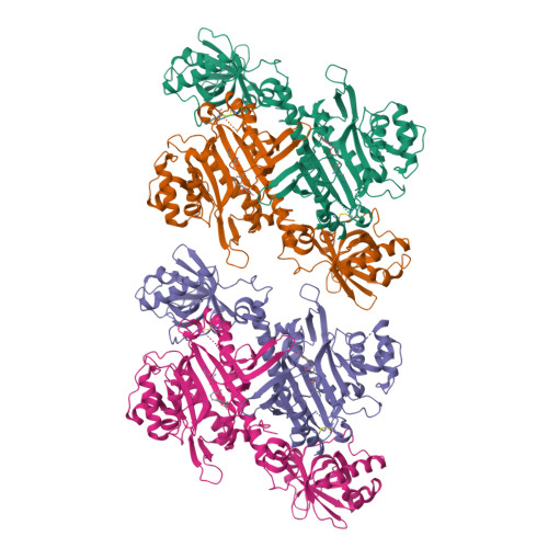
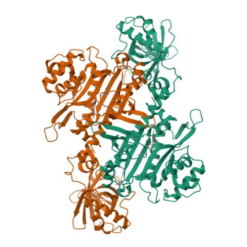
4PG3

Crystal structure of KRS complexed with inhibitor


Experimental Data Snapshot

  • Method: X-RAY DIFFRACTION
  • Resolution: 2.70 Å
  • R-Value Free: 0.247 
  • R-Value Work: 0.193 
  • R-Value Observed: 0.196 

Starting Model: experimental
View more details

wwPDB Validation   3D Report Full Report


Ligand Structure Quality Assessment 


This is version 1.2 of the entry. See complete history


Literature

Structural basis of malaria parasite lysyl-tRNA synthetase inhibition by cladosporin.

Khan, S.Sharma, A.Belrhali, H.Yogavel, M.Sharma, A.

(2014) J Struct Funct Genomics 15: 63-71

  • DOI: https://doi.org/10.1007/s10969-014-9182-1
  • Primary Citation of Related Structures:  
    4PG3

  • PubMed Abstract: 

    Malaria parasites inevitably develop drug resistance to anti-malarials over time. Hence the immediacy for discovering new chemical scaffolds to include in combination malaria drug therapy. The desirable attributes of new chemotherapeutic agents currently include activity against both liver and blood stage malaria parasites. One such recently discovered compound called cladosporin abrogates parasite growth via inhibition of Plasmodium falciparum lysyl-tRNA synthetase (PfKRS), an enzyme central to protein translation. Here, we present crystal structure of ternary PfKRS-lysine-cladosporin (PfKRS-K-C) complex that reveals cladosporin's remarkable ability to mimic the natural substrate adenosine and thereby colonize PfKRS active site. The isocoumarin fragment of cladosporin sandwiches between critical adenine-recognizing residues while its pyran ring fits snugly in the ribose-recognizing cavity. PfKRS-K-C structure highlights ample space within PfKRS active site for further chemical derivatization of cladosporin. Such derivatives may be useful against additional human pathogens that retain high conservation in cladosporin chelating residues within their lysyl-tRNA synthetase.


  • Organizational Affiliation

    Structural and Computational Biology Group, International Centre for Genetic Engineering and Biotechnology (ICGEB), New Delhi, 110067, India.


Macromolecules
Find similar proteins by:  (by identity cutoff)  |  3D Structure
Entity ID: 1
MoleculeChains Sequence LengthOrganismDetailsImage
Lysine--tRNA ligase
A, B, C, D
507Plasmodium falciparum 3D7Mutation(s): 0 
Gene Names: PF13_0262
EC: 6.1.1.6
UniProt
Find proteins for Q8IDJ8 (Plasmodium falciparum (isolate 3D7))
Explore Q8IDJ8 
Go to UniProtKB:  Q8IDJ8
Entity Groups  
Sequence Clusters30% Identity50% Identity70% Identity90% Identity95% Identity100% Identity
UniProt GroupQ8IDJ8
Sequence Annotations
Expand
  • Reference Sequence
Small Molecules
Ligands 2 Unique
IDChains Name / Formula / InChI Key2D Diagram3D Interactions
KRS
Query on KRS

Download Ideal Coordinates CCD File 
F [auth A],
H [auth B],
J [auth C],
L [auth D]
cladosporin
C16 H20 O5
WOMKDMUZNBFXKG-ZWKOPEQDSA-N
LYS
Query on LYS

Download Ideal Coordinates CCD File 
E [auth A],
G [auth B],
I [auth C],
K [auth D]
LYSINE
C6 H15 N2 O2
KDXKERNSBIXSRK-YFKPBYRVSA-O
Experimental Data & Validation

Experimental Data

  • Method: X-RAY DIFFRACTION
  • Resolution: 2.70 Å
  • R-Value Free: 0.247 
  • R-Value Work: 0.193 
  • R-Value Observed: 0.196 
  • Space Group: P 21 21 2
Unit Cell:
Length ( Å )Angle ( ˚ )
a = 297.913α = 90
b = 59.027β = 90
c = 141.457γ = 90
Software Package:
Software NamePurpose
PHENIXrefinement

Structure Validation

View Full Validation Report


Ligand Structure Quality Assessment 


Entry History & Funding Information

Deposition Data


Funding OrganizationLocationGrant Number
Department of BiotechnologyIndiaPR6303

Revision History  (Full details and data files)

  • Version 1.0: 2014-07-16
    Type: Initial release
  • Version 1.1: 2015-03-25
    Changes: Non-polymer description
  • Version 1.2: 2023-09-27
    Changes: Data collection, Database references, Derived calculations, Refinement description, Source and taxonomy