4UNU

MCG - a dimer of lambda variable domains


Experimental Data Snapshot

  • Method: X-RAY DIFFRACTION
  • Resolution: 0.95 Å
  • R-Value Free: 0.130 
  • R-Value Work: 0.107 
  • R-Value Observed: 0.108 

Starting Model: experimental
View more details

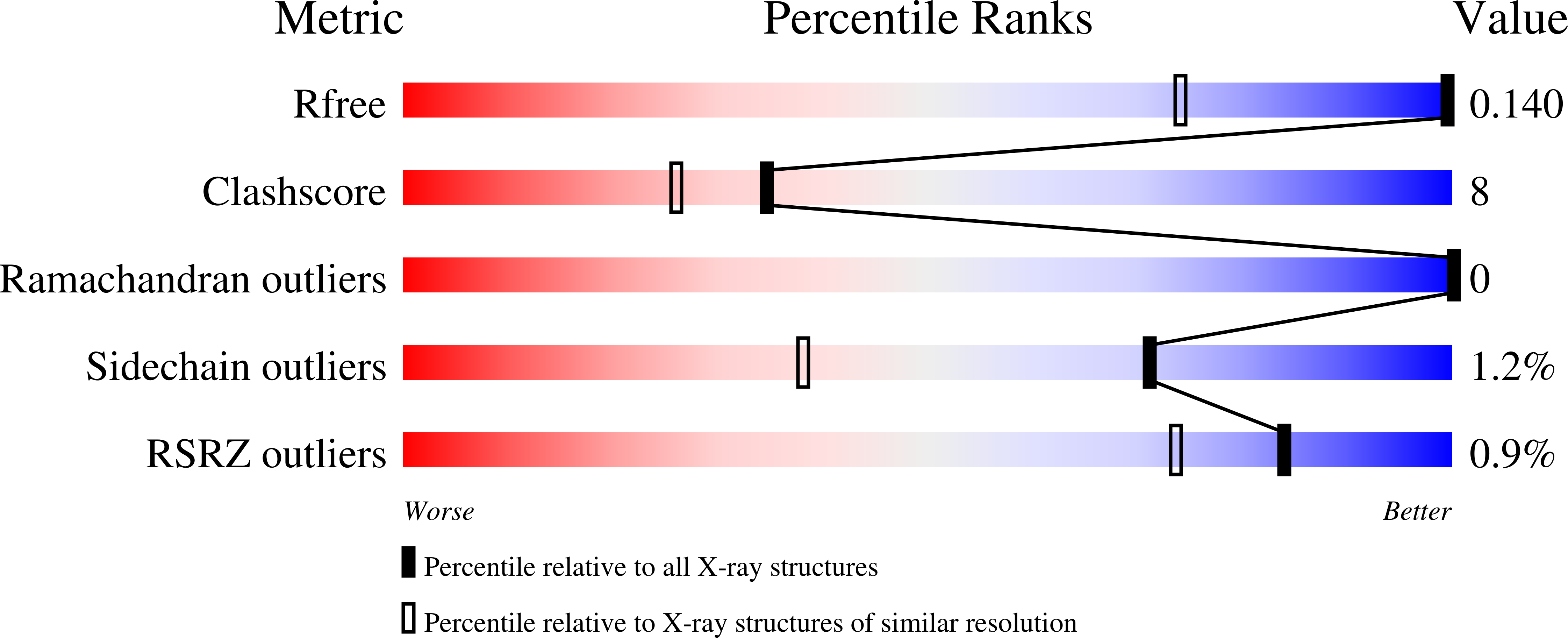
wwPDB Validation   3D Report Full Report


This is version 1.4 of the entry. See complete history


Literature

Formation of Amyloid Fibers by Monomeric Light-Chain Variable Domains.

Brumshtein, B.Esswein, S.R.Landau, M.Ryan, C.M.Whitelegge, J.P.Phillips, M.L.Cascio, D.Sawaya, M.R.Eisenberg, D.S.

(2014) J Biol Chem 289: 27513

  • DOI: https://doi.org/10.1074/jbc.M114.585638
  • Primary Citation of Related Structures:  
    4UNT, 4UNU, 4UNV

  • PubMed Abstract: 

    Systemic light chain amyloidosis is a lethal disease characterized by excess immunoglobulin light chains and light chain fragments composed of variable domains, which aggregate into amyloid fibers. These fibers accumulate and damage organs. Some light chains induce formation of amyloid fibers, whereas others do not, making it unclear what distinguishes amyloid formers from non-formers. One mechanism by which sequence variation may reduce propensity to form amyloid fibers is by shifting the equilibrium toward an amyloid-resistant quaternary structure. Here we identify the monomeric form of the Mcg immunoglobulin light chain variable domain as the quaternary unit required for amyloid fiber assembly. Dimers of Mcg variable domains remain stable and soluble, yet become prone to assemble into amyloid fibers upon disassociation into monomers.


  • Organizational Affiliation

    From the Departments of Biological Chemistry and Chemistry and Biochemistry, Howard Hughes Medical Institute, UCLA-Department of Energy (DOE) Institute for Genomics and Proteomics, UCLA, Los Angeles, California 90095 and.


Macromolecules
Find similar proteins by:  (by identity cutoff)  |  3D Structure
Entity ID: 1
MoleculeChains Sequence LengthOrganismDetailsImage
IG LAMBDA CHAIN V-II REGION MGC
A, B
111Homo sapiensMutation(s): 0 
UniProt & NIH Common Fund Data Resources
Find proteins for P01709 (Homo sapiens)
Explore P01709 
Go to UniProtKB:  P01709
GTEx:  ENSG00000278196 
Entity Groups  
Sequence Clusters30% Identity50% Identity70% Identity90% Identity95% Identity100% Identity
UniProt GroupP01709
Sequence Annotations
Expand
  • Reference Sequence
Experimental Data & Validation

Experimental Data

  • Method: X-RAY DIFFRACTION
  • Resolution: 0.95 Å
  • R-Value Free: 0.130 
  • R-Value Work: 0.107 
  • R-Value Observed: 0.108 
  • Space Group: P 1 21 1
Unit Cell:
Length ( Å )Angle ( ˚ )
a = 41.34α = 90
b = 34.12β = 104.94
c = 63.18γ = 90
Software Package:
Software NamePurpose
REFMACrefinement
XDSdata reduction
XDSdata scaling
PHASERphasing

Structure Validation

View Full Validation Report


Entry History 

Deposition Data

Revision History  (Full details and data files)

  • Version 1.0: 2014-08-27
    Type: Initial release
  • Version 1.1: 2014-09-03
    Changes: Database references
  • Version 1.2: 2014-10-15
    Changes: Database references
  • Version 1.3: 2017-09-13
    Changes: Data collection
  • Version 1.4: 2024-01-10
    Changes: Data collection, Database references, Derived calculations, Other, Refinement description