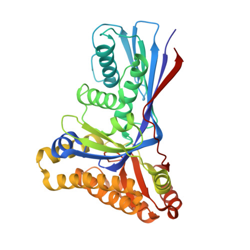
4UTG | pdb_00004utg

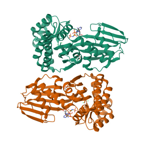
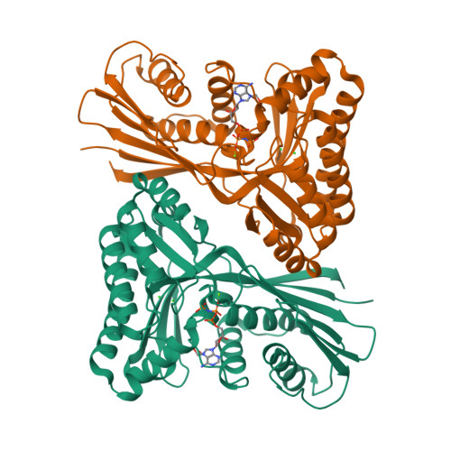
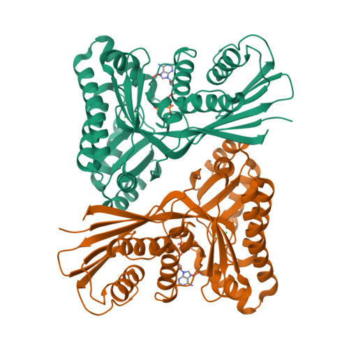
Burkholderia pseudomallei heptokinase WcbL,AMPPNP (ATP analogue) complex.


Experimental Data Snapshot

  • Method: X-RAY DIFFRACTION
  • Resolution: 1.93 Å
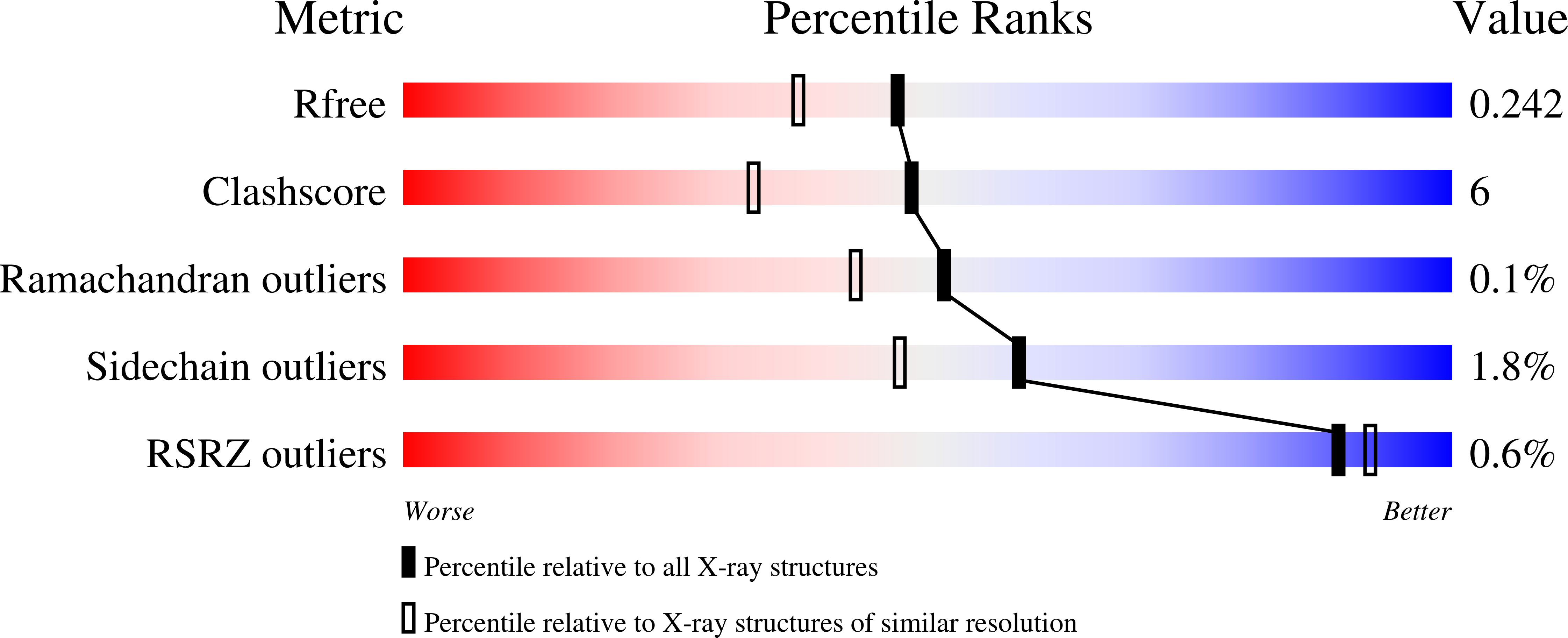
  • R-Value Free: 
    0.242 (Depositor), 0.240 (DCC) 
  • R-Value Work: 
    0.194 (Depositor), 0.190 (DCC) 
  • R-Value Observed: 
    0.196 (Depositor) 

wwPDB Validation   3D Report Full Report


Ligand Structure Quality Assessment 

Created with Raphaël 2.3.0Worse 01 BetterLigand structure goodness of fit to experimental dataBest fitted ANPClick on this verticalbar to view details

This is version 1.2 of the entry. See complete history


Literature

Unraveling the B.Pseudomallei Heptokinase Wcbl: From Structure to Drug Discovery.

Vivoli, M.Isupov, M.N.Nicholas, R.Hill, A.Scott, A.E.Kosma, P.Prior, J.L.Harmer, N.J.

(2015) Chem Biol 22: 1622

  • DOI: https://doi.org/10.1016/j.chembiol.2015.10.015
  • Primary Citation of Related Structures:  
    4USK, 4USM, 4UT4, 4UTG

  • PubMed Abstract: 

    Gram-negative bacteria utilize heptoses as part of their repertoire of extracellular polysaccharide virulence determinants. Disruption of heptose biosynthesis offers an attractive target for novel antimicrobials. A critical step in the synthesis of heptoses is their 1-O phosphorylation, mediated by kinases such as HldE or WcbL. Here, we present the structure of WcbL from Burkholderia pseudomallei. We report that WcbL operates through a sequential ordered Bi-Bi mechanism, loading the heptose first and then ATP. We show that dimeric WcbL binds ATP anti-cooperatively in the absence of heptose, and cooperatively in its presence. Modeling of WcbL suggests that heptose binding causes an elegant switch in the hydrogen-bonding network, facilitating the binding of a second ATP molecule. Finally, we screened a library of drug-like fragments, identifying hits that potently inhibit WcbL. Our results provide a novel mechanism for control of substrate binding and emphasize WcbL as an attractive anti-microbial target for Gram-negative bacteria.


  • Organizational Affiliation

    Department of Biosciences, University of Exeter, Henry Wellcome Building, Stocker Road, Exeter EX4 4QD, UK.


Macromolecules
Find similar proteins by:  (by identity cutoff)  |  3D Structure
Entity ID: 1
MoleculeChains Sequence LengthOrganismDetailsImage
SUGAR KINASE
A, B
346Burkholderia pseudomallei K96243Mutation(s): 3 
EC: 2.7.1.167
UniProt
Find proteins for H7C745 (Burkholderia pseudomallei (strain K96243))
Explore H7C745 
Go to UniProtKB:  H7C745
Entity Groups  
Sequence Clusters30% Identity50% Identity70% Identity90% Identity95% Identity100% Identity
UniProt GroupH7C745
Sequence Annotations
Expand
  • Reference Sequence
Small Molecules
Ligands 4 Unique
IDChains Name / Formula / InChI Key2D Diagram3D Interactions
ANP
Query on ANP

Download Ideal Coordinates CCD File 
C [auth A],
H [auth B]
PHOSPHOAMINOPHOSPHONIC ACID-ADENYLATE ESTER
C10 H17 N6 O12 P3
PVKSNHVPLWYQGJ-KQYNXXCUSA-N
EDO
Query on EDO

Download Ideal Coordinates CCD File 
G [auth A],
L [auth B]
1,2-ETHANEDIOL
C2 H6 O2
LYCAIKOWRPUZTN-UHFFFAOYSA-N
CL
Query on CL

Download Ideal Coordinates CCD File 
E [auth A],
F [auth A],
J [auth B],
K [auth B]
CHLORIDE ION
Cl
VEXZGXHMUGYJMC-UHFFFAOYSA-M
MG
Query on MG

Download Ideal Coordinates CCD File 
D [auth A],
I [auth B]
MAGNESIUM ION
Mg
JLVVSXFLKOJNIY-UHFFFAOYSA-N
Experimental Data & Validation

Experimental Data

  • Method: X-RAY DIFFRACTION
  • Resolution: 1.93 Å
  • R-Value Free:  0.242 (Depositor), 0.240 (DCC) 
  • R-Value Work:  0.194 (Depositor), 0.190 (DCC) 
  • R-Value Observed: 0.196 (Depositor) 
Space Group: C 2 2 21
Unit Cell:
Length ( Å )Angle ( ˚ )
a = 101.44α = 90
b = 115.02β = 90
c = 166.78γ = 90
Software Package:
Software NamePurpose
xia2data reduction
XDSdata reduction
Aimlessdata scaling
SHELXphasing
ARP/wARPphasing
REFMACrefinement

Structure Validation

View Full Validation Report


Ligand Structure Quality Assessment 

Created with Raphaël 2.3.0Worse 01 BetterLigand structure goodness of fit to experimental dataBest fitted ANPClick on this verticalbar to view details

Entry History 

Deposition Data

Revision History  (Full details and data files)

  • Version 1.0: 2016-01-13
    Type: Initial release
  • Version 1.1: 2017-06-28
    Changes: Refinement description
  • Version 1.2: 2024-05-08
    Changes: Data collection, Database references, Derived calculations, Other