4WT6 | pdb_00004wt6

Crystal structure of human Pim-1 kinase in complex with a thiadiazolamine-indole inhibitor.


Experimental Data Snapshot

  • Method: X-RAY DIFFRACTION
  • Resolution: 2.30 Å
  • R-Value Free: 
    0.211 (Depositor), 0.220 (DCC) 
  • R-Value Work: 
    0.176 (Depositor), 0.180 (DCC) 
  • R-Value Observed: 
    0.178 (Depositor) 

wwPDB Validation   3D Report Full Report


Ligand Structure Quality Assessment 

Created with Raphaël 2.3.0Worse 01 BetterLigand structure goodness of fit to experimental dataBest fitted 3U6Click on this verticalbar to view details

This is version 1.1 of the entry. See complete history


Literature

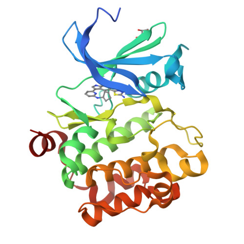
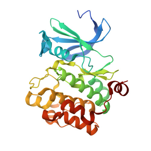
Discovery of 5-(1H-indol-5-yl)-1,3,4-thiadiazol-2-amines as potent PIM inhibitors.

Wu, B.Wang, H.L.Cee, V.J.Lanman, B.A.Nixey, T.Pettus, L.Reed, A.B.Wurz, R.P.Guerrero, N.Sastri, C.Winston, J.Lipford, J.R.Lee, M.R.Mohr, C.Andrews, K.L.Tasker, A.S.

(2015) Bioorg Med Chem Lett 25: 775-780

  • DOI: https://doi.org/10.1016/j.bmcl.2014.12.091
  • Primary Citation of Related Structures:  
    4WSY, 4WT6

  • PubMed Abstract: 

    PIM kinases are a family of Ser/Thr kinases that are implicated in tumorigenesis. The discovery of a new class of PIM inhibitors, 5-(1H-indol-5-yl)-1,3,4-thiadiazol-2-amines, is discussed with optimized compounds showing excellent potency against all three PIM isoforms.


  • Organizational Affiliation

    Department of Therapeutic Discovery, Amgen Inc., One Amgen Center Drive, Thousand Oaks, CA 91320-1799, USA. Electronic address: binw@amgen.com.


Macromolecules
Find similar proteins by:  (by identity cutoff)  |  3D Structure
Entity ID: 1
MoleculeChains Sequence LengthOrganismDetailsImage
Serine/threonine-protein kinase pim-1286Homo sapiensMutation(s): 0 
Gene Names: PIM1
EC: 2.7.11.1
UniProt & NIH Common Fund Data Resources
Find proteins for P11309 (Homo sapiens)
Explore P11309 
Go to UniProtKB:  P11309
PHAROS:  P11309
GTEx:  ENSG00000137193 
Entity Groups  
Sequence Clusters30% Identity50% Identity70% Identity90% Identity95% Identity100% Identity
UniProt GroupP11309
Sequence Annotations
Expand
  • Reference Sequence
Small Molecules
Ligands 2 Unique
IDChains Name / Formula / InChI Key2D Diagram3D Interactions
3U6
Query on 3U6

Download Ideal Coordinates CCD File 
B [auth A]6-[5-(5-amino-1,3,4-thiadiazol-2-yl)-1H-indol-3-yl]-N-cyclopentylpyridin-2-amine
C20 H20 N6 S
LYSVYDGAEVIKAF-UHFFFAOYSA-N
GOL
Query on GOL

Download Ideal Coordinates CCD File 
C [auth A]GLYCEROL
C3 H8 O3
PEDCQBHIVMGVHV-UHFFFAOYSA-N
Binding Affinity Annotations 
IDSourceBinding Affinity
3U6 BindingDB:  4WT6 IC50: min: 12, max: 111 (nM) from 3 assay(s)
Experimental Data & Validation

Experimental Data

  • Method: X-RAY DIFFRACTION
  • Resolution: 2.30 Å
  • R-Value Free:  0.211 (Depositor), 0.220 (DCC) 
  • R-Value Work:  0.176 (Depositor), 0.180 (DCC) 
  • R-Value Observed: 0.178 (Depositor) 
Space Group: P 65
Unit Cell:
Length ( Å )Angle ( ˚ )
a = 98.184α = 90
b = 98.184β = 90
c = 80.8γ = 120
Software Package:
Software NamePurpose
REFMACrefinement
SCALEPACKdata scaling
HKL-2000data reduction
MOLREPphasing

Structure Validation

View Full Validation Report


Ligand Structure Quality Assessment 

Created with Raphaël 2.3.0Worse 01 BetterLigand structure goodness of fit to experimental dataBest fitted 3U6Click on this verticalbar to view details

Entry History 

Deposition Data

  • Released Date: 2015-02-11 
  • Deposition Author(s): Mohr, C.

Revision History  (Full details and data files)

  • Version 1.0: 2015-02-11
    Type: Initial release
  • Version 1.1: 2023-12-27
    Changes: Data collection, Database references, Derived calculations, Other, Source and taxonomy, Structure summary