4X1R

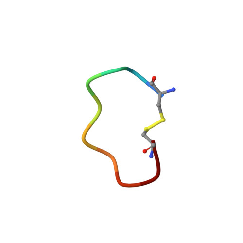
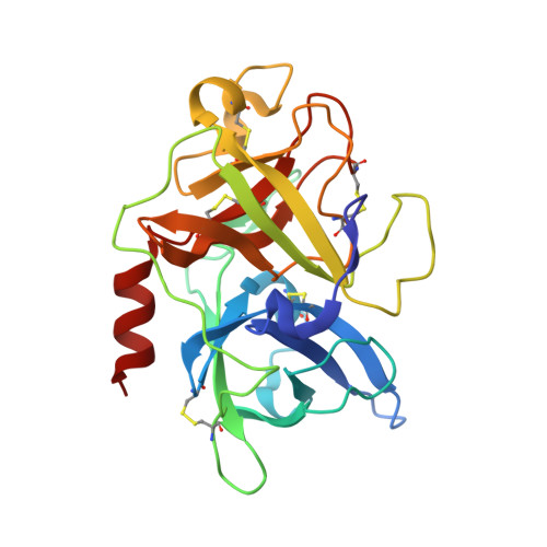
The crystal structure of mupain-1-12 in complex with murinised human uPA at pH7.4


Experimental Data Snapshot

  • Method: X-RAY DIFFRACTION
  • Resolution: 2.10 Å
  • R-Value Free: 0.273 
  • R-Value Work: 0.210 
  • R-Value Observed: 0.213 

Starting Model: experimental
View more details

wwPDB Validation   3D Report Full Report


This is version 1.1 of the entry. See complete history


Literature

A cyclic peptidic serine protease inhibitor: increasing affinity by increasing peptide flexibility.

Zhao, B.Xu, P.Jiang, L.Paaske, B.Kromann-Hansen, T.Jensen, J.K.Srensen, H.P.Liu, Z.Nielsen, J.T.Christensen, A.Hosseini, M.Srensen, K.K.Nielsen, N.C.Jensen, K.J.Huang, M.Andreasen, P.A.

(2014) PLoS One 9: e115872-e115872

  • DOI: https://doi.org/10.1371/journal.pone.0115872
  • Primary Citation of Related Structures:  
    4X1N, 4X1Q, 4X1R, 4X1S

  • PubMed Abstract: 

    Peptides are attracting increasing interest as protease inhibitors. Here, we demonstrate a new inhibitory mechanism and a new type of exosite interactions for a phage-displayed peptide library-derived competitive inhibitor, mupain-1 (CPAYSRYLDC), of the serine protease murine urokinase-type plasminogen activator (uPA). We used X-ray crystal structure analysis, site-directed mutagenesis, liquid state NMR, surface plasmon resonance analysis, and isothermal titration calorimetry and wild type and engineered variants of murine and human uPA. We demonstrate that Arg6 inserts into the S1 specificity pocket, its carbonyl group aligning improperly relative to Ser195 and the oxyanion hole, explaining why the peptide is an inhibitor rather than a substrate. Substitution of the P1 Arg with novel unnatural Arg analogues with aliphatic or aromatic ring structures led to an increased affinity, depending on changes in both P1 - S1 and exosite interactions. Site-directed mutagenesis showed that exosite interactions, while still supporting high affinity binding, differed substantially between different uPA variants. Surprisingly, high affinity binding was facilitated by Ala-substitution of Asp9 of the peptide, in spite of a less favorable binding entropy and loss of a polar interaction. We conclude that increased flexibility of the peptide allows more favorable exosite interactions, which, in combination with the use of novel Arg analogues as P1 residues, can be used to manipulate the affinity and specificity of this peptidic inhibitor, a concept different from conventional attempts at improving inhibitor affinity by reducing the entropic burden.


  • Organizational Affiliation

    Danish-Chinese Centre for Proteases and Cancer, Fujian Institute of Research on the Structure of Matter, Chinese Academy of Sciences, Fuzhou, China.


Macromolecules

Find similar proteins by:  Sequence   |   3D Structure  

Entity ID: 1
MoleculeChains Sequence LengthOrganismDetailsImage
mupain-1-12A [auth P]10synthetic constructMutation(s): 0 
Entity Groups  
Sequence Clusters30% Identity50% Identity70% Identity90% Identity95% Identity100% Identity
Sequence Annotations
Expand
  • Reference Sequence
Find similar proteins by:  (by identity cutoff)  |  3D Structure
Entity ID: 2
MoleculeChains Sequence LengthOrganismDetailsImage
Urokinase-type plasminogen activatorB [auth U]247Homo sapiensMutation(s): 3 
Gene Names: PLAU
EC: 3.4.21.73
UniProt & NIH Common Fund Data Resources
Find proteins for P00749 (Homo sapiens)
Explore P00749 
Go to UniProtKB:  P00749
PHAROS:  P00749
GTEx:  ENSG00000122861 
Entity Groups  
Sequence Clusters30% Identity50% Identity70% Identity90% Identity95% Identity100% Identity
UniProt GroupP00749
Sequence Annotations
Expand
  • Reference Sequence
Small Molecules
Ligands 1 Unique
IDChains Name / Formula / InChI Key2D Diagram3D Interactions
PL0
Query on PL0

Download Ideal Coordinates CCD File 
C [auth P]1-phenylguanidine
C7 H9 N3
QRJZGVVKGFIGLI-UHFFFAOYSA-N
Binding Affinity Annotations 
IDSourceBinding Affinity
PL0 BindingDB:  4X1R Ki: min: 2.06e+4, max: 2.06e+4 (nM) from 2 assay(s)
IC50: 2.00e+4 (nM) from 1 assay(s)
Experimental Data & Validation

Experimental Data

  • Method: X-RAY DIFFRACTION
  • Resolution: 2.10 Å
  • R-Value Free: 0.273 
  • R-Value Work: 0.210 
  • R-Value Observed: 0.213 
  • Space Group: H 3
Unit Cell:
Length ( Å )Angle ( ˚ )
a = 121.177α = 90
b = 121.177β = 90
c = 42.368γ = 120
Software Package:
Software NamePurpose
REFMACrefinement
Cootmodel building

Structure Validation

View Full Validation Report


Entry History 

Deposition Data

Revision History  (Full details and data files)

  • Version 1.0: 2015-03-25
    Type: Initial release
  • Version 1.1: 2023-11-08
    Changes: Data collection, Database references, Derived calculations, Refinement description, Source and taxonomy