4YCI

non-latent pro-bone morphogenetic protein 9


Experimental Data Snapshot

  • Method: X-RAY DIFFRACTION
  • Resolution: 3.25 Å
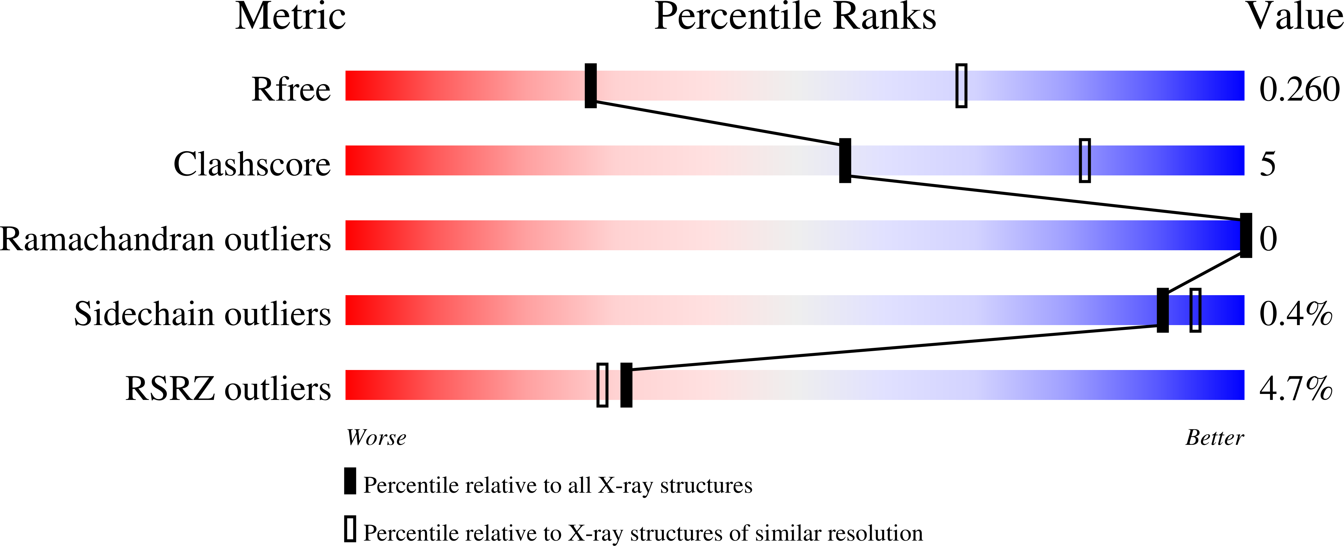
  • R-Value Free: 0.260 
  • R-Value Work: 0.240 
  • R-Value Observed: 0.241 

Starting Model: experimental
View more details

wwPDB Validation   3D Report Full Report


Ligand Structure Quality Assessment 


This is version 2.2 of the entry. See complete history


Literature

Structure of bone morphogenetic protein 9 procomplex.

Mi, L.Z.Brown, C.T.Gao, Y.Tian, Y.Le, V.Q.Walz, T.Springer, T.A.

(2015) Proc Natl Acad Sci U S A 112: 3710-3715

  • DOI: https://doi.org/10.1073/pnas.1501303112
  • Primary Citation of Related Structures:  
    4YCG, 4YCI

  • PubMed Abstract: 

    Bone morphogenetic proteins (BMPs) belong to the TGF-β family, whose 33 members regulate multiple aspects of morphogenesis. TGF-β family members are secreted as procomplexes containing a small growth factor dimer associated with two larger prodomains. As isolated procomplexes, some members are latent, whereas most are active; what determines these differences is unknown. Here, studies on pro-BMP structures and binding to receptors lead to insights into mechanisms that regulate latency in the TGF-β family and into the functions of their highly divergent prodomains. The observed open-armed, nonlatent conformation of pro-BMP9 and pro-BMP7 contrasts with the cross-armed, latent conformation of pro-TGF-β1. Despite markedly different arm orientations in pro-BMP and pro-TGF-β, the arm domain of the prodomain can similarly associate with the growth factor, whereas prodomain elements N- and C-terminal to the arm associate differently with the growth factor and may compete with one another to regulate latency and stepwise displacement by type I and II receptors. Sequence conservation suggests that pro-BMP9 can adopt both cross-armed and open-armed conformations. We propose that interactors in the matrix stabilize a cross-armed pro-BMP conformation and regulate transition between cross-armed, latent and open-armed, nonlatent pro-BMP conformations.


  • Organizational Affiliation

    Department of Biological Chemistry and Molecular Pharmacology, Harvard Medical School, Boston, MA 02115; Program in Cellular and Molecular Medicine, Children's Hospital Boston, Boston, MA 02115; Department of Molecular and Structural Biology, School of Life Sciences, Tianjin University, Tianjin 300072, China;


Macromolecules
Find similar proteins by:  (by identity cutoff)  |  3D Structure
Entity ID: 1
MoleculeChains Sequence LengthOrganismDetailsImage
Bone Morphogenetic Protein 9 Growth Factor Domain
A, B
296Mus musculusMutation(s): 0 
Gene Names: Gdf2Bmp9
UniProt & NIH Common Fund Data Resources
Find proteins for Q9WV56 (Mus musculus)
Explore Q9WV56 
Go to UniProtKB:  Q9WV56
IMPC:  MGI:1321394
Entity Groups  
Sequence Clusters30% Identity50% Identity70% Identity90% Identity95% Identity100% Identity
UniProt GroupQ9WV56
Glycosylation
Glycosylation Sites: 1Go to GlyGen: Q9WV56-1
Sequence Annotations
Expand
  • Reference Sequence
Find similar proteins by:  (by identity cutoff)  |  3D Structure
Entity ID: 2
MoleculeChains Sequence LengthOrganismDetailsImage
Bone Morphogenetic Protein 9 Prodomain
C, D
110Homo sapiensMutation(s): 0 
Gene Names: GDF2BMP9
UniProt & NIH Common Fund Data Resources
Find proteins for Q9UK05 (Homo sapiens)
Explore Q9UK05 
Go to UniProtKB:  Q9UK05
PHAROS:  Q9UK05
GTEx:  ENSG00000263761 
Entity Groups  
Sequence Clusters30% Identity50% Identity70% Identity90% Identity95% Identity100% Identity
UniProt GroupQ9UK05
Sequence Annotations
Expand
  • Reference Sequence
Oligosaccharides

Help

Entity ID: 3
MoleculeChains Length2D Diagram Glycosylation3D Interactions
2-acetamido-2-deoxy-beta-D-glucopyranose-(1-4)-2-acetamido-2-deoxy-beta-D-glucopyranose
E
2N-Glycosylation
Glycosylation Resources
GlyTouCan:  G42666HT
GlyCosmos:  G42666HT
GlyGen:  G42666HT
Small Molecules
Ligands 2 Unique
IDChains Name / Formula / InChI Key2D Diagram3D Interactions
NAG
Query on NAG

Download Ideal Coordinates CCD File 
F [auth A]2-acetamido-2-deoxy-beta-D-glucopyranose
C8 H15 N O6
OVRNDRQMDRJTHS-FMDGEEDCSA-N
ZN
Query on ZN

Download Ideal Coordinates CCD File 
G [auth A]
H [auth A]
I [auth A]
J [auth A]
K [auth B]
G [auth A],
H [auth A],
I [auth A],
J [auth A],
K [auth B],
L [auth B],
M [auth B],
N [auth B],
O [auth C],
P [auth C]
ZINC ION
Zn
PTFCDOFLOPIGGS-UHFFFAOYSA-N
Experimental Data & Validation

Experimental Data

  • Method: X-RAY DIFFRACTION
  • Resolution: 3.25 Å
  • R-Value Free: 0.260 
  • R-Value Work: 0.240 
  • R-Value Observed: 0.241 
  • Space Group: P 32 2 1
Unit Cell:
Length ( Å )Angle ( ˚ )
a = 120.28α = 90
b = 120.28β = 90
c = 221.31γ = 120
Software Package:
Software NamePurpose
PHENIXrefinement
XSCALEdata scaling
PDB_EXTRACTdata extraction
XDSdata reduction
PHENIXphasing

Structure Validation

View Full Validation Report


Ligand Structure Quality Assessment 


Entry History 

Deposition Data

Revision History  (Full details and data files)

  • Version 1.0: 2015-03-04
    Type: Initial release
  • Version 1.1: 2015-03-11
    Changes: Structure summary
  • Version 1.2: 2015-03-25
    Changes: Database references
  • Version 1.3: 2015-04-01
    Changes: Database references
  • Version 1.4: 2015-08-26
    Changes: Structure summary
  • Version 2.0: 2020-07-29
    Type: Remediation
    Reason: Carbohydrate remediation
    Changes: Atomic model, Data collection, Derived calculations, Structure summary
  • Version 2.1: 2023-09-27
    Changes: Data collection, Database references, Refinement description, Structure summary
  • Version 2.2: 2024-10-23
    Changes: Structure summary