4ZS9

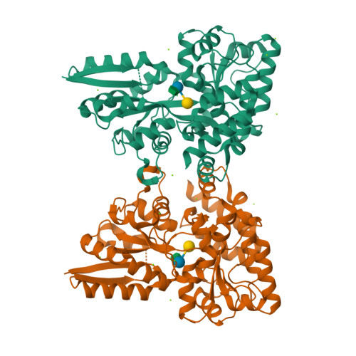
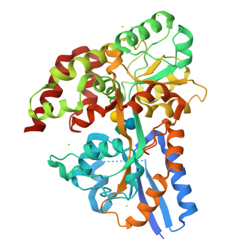
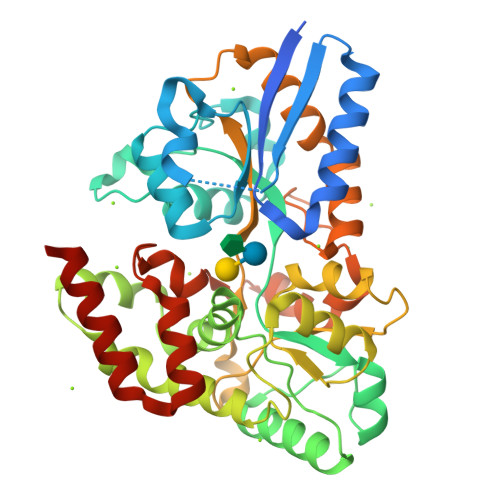
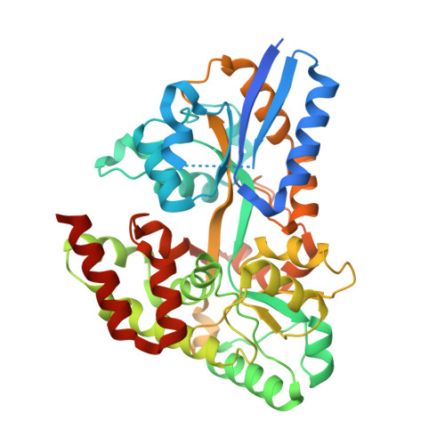
Raffinose and panose binding protein from Bifidobacterium animalis subsp. lactis Bl-04, bound with raffinose


Experimental Data Snapshot

  • Method: X-RAY DIFFRACTION
  • Resolution: 1.37 Å
  • R-Value Free: 0.167 
  • R-Value Work: 0.132 
  • R-Value Observed: 0.134 

Starting Model: experimental
View more details

wwPDB Validation   3D Report Full Report


This is version 2.1 of the entry. See complete history


Literature

An ATP Binding Cassette Transporter Mediates the Uptake of alpha-(1,6)-Linked Dietary Oligosaccharides in Bifidobacterium and Correlates with Competitive Growth on These Substrates.

Ejby, M.Fredslund, F.Andersen, J.M.Vujicic Zagar, A.Henriksen, J.R.Andersen, T.L.Svensson, B.Slotboom, D.J.Abou Hachem, M.

(2016) J Biol Chem 291: 20220-20231

  • DOI: https://doi.org/10.1074/jbc.M116.746529
  • Primary Citation of Related Structures:  
    4ZS9, 4ZZA, 4ZZE

  • PubMed Abstract: 

    The molecular details and impact of oligosaccharide uptake by distinct human gut microbiota (HGM) are currently not well understood. Non-digestible dietary galacto- and gluco-α-(1,6)-oligosaccharides from legumes and starch, respectively, are preferentially fermented by mainly bifidobacteria and lactobacilli in the human gut. Here we show that the solute binding protein (BlG16BP) associated with an ATP binding cassette (ABC) transporter from the probiotic Bifidobacterium animalis subsp. lactis Bl-04 binds α-(1,6)-linked glucosides and galactosides of varying size, linkage, and monosaccharide composition with preference for the trisaccharides raffinose and panose. This preference is also reflected in the α-(1,6)-galactoside uptake profile of the bacterium. Structures of BlG16BP in complex with raffinose and panose revealed the basis for the remarkable ligand binding plasticity of BlG16BP, which recognizes the non-reducing α-(1,6)-diglycoside in its ligands. BlG16BP homologues occur predominantly in bifidobacteria and a few Firmicutes but lack in other HGMs. Among seven bifidobacterial taxa, only those possessing this transporter displayed growth on α-(1,6)-glycosides. Competition assays revealed that the dominant HGM commensal Bacteroides ovatus was out-competed by B. animalis subsp. lactis Bl-04 in mixed cultures growing on raffinose, the preferred ligand for the BlG16BP. By comparison, B. ovatus mono-cultures grew very efficiently on this trisaccharide. These findings suggest that the ABC-mediated uptake of raffinose provides an important competitive advantage, particularly against dominant Bacteroides that lack glycan-specific ABC-transporters. This novel insight highlights the role of glycan transport in defining the metabolic specialization of gut bacteria.


  • Organizational Affiliation

    From the Protein Glycoscience and Biotechnology, Department of Bioengineering, Elektrovej, Building 375.


Macromolecules
Find similar proteins by:  (by identity cutoff)  |  3D Structure
Entity ID: 1
MoleculeChains Sequence LengthOrganismDetailsImage
Sugar binding protein of ABC transporter system
A, B
413Bifidobacterium animalis subsp. lactis Bl-04Mutation(s): 0 
Gene Names: Balac_1599
UniProt
Find proteins for A0A1A9TA69 (Bifidobacterium animalis subsp. lactis (strain Bl-04 / DGCC2908 / RB 4825 / SD5219))
Explore A0A1A9TA69 
Go to UniProtKB:  A0A1A9TA69
Entity Groups  
Sequence Clusters30% Identity50% Identity70% Identity90% Identity95% Identity100% Identity
UniProt GroupA0A1A9TA69
Sequence Annotations
Expand
  • Reference Sequence
Oligosaccharides

Help

Entity ID: 2
MoleculeChains Length2D Diagram Glycosylation3D Interactions
alpha-D-galactopyranose-(1-6)-alpha-D-glucopyranose-(1-2)-beta-D-fructofuranose
C, D
3N/A
Glycosylation Resources
GlyTouCan:  G40178VU
GlyCosmos:  G40178VU
Small Molecules
Ligands 2 Unique
IDChains Name / Formula / InChI Key2D Diagram3D Interactions
CL
Query on CL

Download Ideal Coordinates CCD File 
L [auth A],
M [auth A],
V [auth B],
W [auth B]
CHLORIDE ION
Cl
VEXZGXHMUGYJMC-UHFFFAOYSA-M
MG
Query on MG

Download Ideal Coordinates CCD File 
E [auth A]
F [auth A]
G [auth A]
H [auth A]
I [auth A]
E [auth A],
F [auth A],
G [auth A],
H [auth A],
I [auth A],
J [auth A],
K [auth A],
N [auth B],
O [auth B],
P [auth B],
Q [auth B],
R [auth B],
S [auth B],
T [auth B],
U [auth B]
MAGNESIUM ION
Mg
JLVVSXFLKOJNIY-UHFFFAOYSA-N
Biologically Interesting Molecules (External Reference) 1 Unique
Experimental Data & Validation

Experimental Data

  • Method: X-RAY DIFFRACTION
  • Resolution: 1.37 Å
  • R-Value Free: 0.167 
  • R-Value Work: 0.132 
  • R-Value Observed: 0.134 
  • Space Group: P 21 21 21
Unit Cell:
Length ( Å )Angle ( ˚ )
a = 55.54α = 90
b = 90.82β = 90
c = 146.75γ = 90
Software Package:
Software NamePurpose
xia2data reduction
XSCALEdata scaling
PHASERphasing
PHENIXrefinement
PDB_EXTRACTdata extraction

Structure Validation

View Full Validation Report


Entry History & Funding Information

Deposition Data


Funding OrganizationLocationGrant Number
Denmark--

Revision History  (Full details and data files)

  • Version 1.0: 2016-06-29
    Type: Initial release
  • Version 1.1: 2017-08-16
    Changes: Database references
  • Version 2.0: 2020-07-29
    Type: Remediation
    Reason: Carbohydrate remediation
    Changes: Atomic model, Data collection, Derived calculations, Non-polymer description, Structure summary
  • Version 2.1: 2024-01-10
    Changes: Data collection, Database references, Refinement description, Structure summary