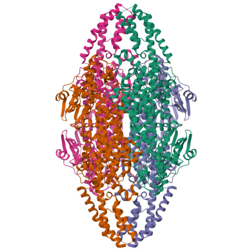
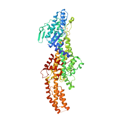
4C5R

Structural Investigations into the Stereochemistry and Activity of a Phenylalanine-2,3-Aminomutase from Taxus chinensis


Experimental Data Snapshot

  • Method: X-RAY DIFFRACTION
  • Resolution: 2.14 Å
  • R-Value Free: 0.210 
  • R-Value Work: 0.172 
  • R-Value Observed: 0.174 

Starting Model: experimental
View more details

wwPDB Validation   3D Report Full Report


Ligand Structure Quality Assessment 


This is version 2.2 of the entry. See complete history


Literature

Structural Investigations Into the Stereochemistry and Activity of a Phenylalanine-2,3-Aminomutase from Taxus Chinensis.

Wybenga, G.G.Szymanski, W.Wu, B.Feringa, B.L.Janssen, D.B.Dijkstra, B.W.

(2014) Biochemistry 53: 3187

  • DOI: https://doi.org/10.1021/bi500187a
  • Primary Citation of Related Structures:  
    4C5R, 4C5S, 4C5U, 4C6G, 4CQ5

  • PubMed Abstract: 

    Phenylalanine-2,3-aminomutase (PAM) from Taxus chinensis, a 4-methylidene-imidazole-5-one (MIO)-dependent enzyme, catalyzes the reversible conversion of (S)-α-phenylalanine into (R)-β-phenylalanine via trans-cinnamic acid. The enzyme also catalyzes the direct addition of ammonia to trans-cinnamic acid, a reaction that can be used for the preparation of β-amino acids, which occur as frequent constituents of bioactive compounds. Different hypotheses have been formulated to explain the stereochemistry of the PAM-catalyzed reaction, but structural evidence for these hypotheses is lacking. Furthermore, it remains unclear how the PAM MIO group is formed from the three-amino acid (A-S-G) sequence motif. For these reasons, we elucidated PAM three-dimensional (3D) structures with a bound (R)-β-phenylalanine analogue and with bound trans-cinnamic acid. In addition, 3D structures of the (inactive) Y322A and N231A mutants of PAM were elucidated, which were found to be MIO-less. We conclude that the stereochemistry of the PAM-catalyzed reaction originates from the enzyme's ability to bind trans-cinnamic acid in two different orientations, with either the si,si face or the re,re face directed toward the MIO group, as evidenced by two distinct carboxylate binding modes. The results also suggest that the N231 side chain promotes MIO group formation by increasing the nucleophilicity of the G177 N atom through acidification of the amide proton.


  • Organizational Affiliation

    Laboratory of Biophysical Chemistry, University of Groningen , Nijenborgh 7, 9747 AG Groningen, The Netherlands.


Macromolecules
Find similar proteins by:  (by identity cutoff)  |  3D Structure
Entity ID: 1
MoleculeChains Sequence LengthOrganismDetailsImage
PHENYLALANINE AMMONIA-LYASE
A, B, C, D
705Taxus chinensisMutation(s): 1 
EC: 4.3.1.24 (PDB Primary Data), 5.4.3.10 (UniProt)
UniProt
Find proteins for Q68G84 (Taxus chinensis)
Explore Q68G84 
Go to UniProtKB:  Q68G84
Entity Groups  
Sequence Clusters30% Identity50% Identity70% Identity90% Identity95% Identity100% Identity
UniProt GroupQ68G84
Sequence Annotations
Expand
  • Reference Sequence
Experimental Data & Validation

Experimental Data

  • Method: X-RAY DIFFRACTION
  • Resolution: 2.14 Å
  • R-Value Free: 0.210 
  • R-Value Work: 0.172 
  • R-Value Observed: 0.174 
  • Space Group: P 1 21 1
Unit Cell:
Length ( Å )Angle ( ˚ )
a = 99.395α = 90
b = 145.477β = 99.54
c = 99.564γ = 90
Software Package:
Software NamePurpose
REFMACrefinement
XDSdata reduction
SCALAdata scaling

Structure Validation

View Full Validation Report


Ligand Structure Quality Assessment 


Entry History 

Deposition Data

Revision History  (Full details and data files)

  • Version 1.0: 2014-05-07
    Type: Initial release
  • Version 1.1: 2014-05-14
    Changes: Database references
  • Version 1.2: 2014-06-04
    Changes: Database references
  • Version 1.3: 2019-10-23
    Changes: Data collection, Database references, Derived calculations, Other
  • Version 2.0: 2023-11-15
    Changes: Atomic model, Data collection, Database references, Derived calculations
  • Version 2.1: 2023-12-20
    Changes: Refinement description
  • Version 2.2: 2024-11-13
    Changes: Structure summary