5AFM

alpha7-AChBP in complex with lobeline and fragment 4


Experimental Data Snapshot

  • Method: X-RAY DIFFRACTION
  • Resolution: 2.85 Å
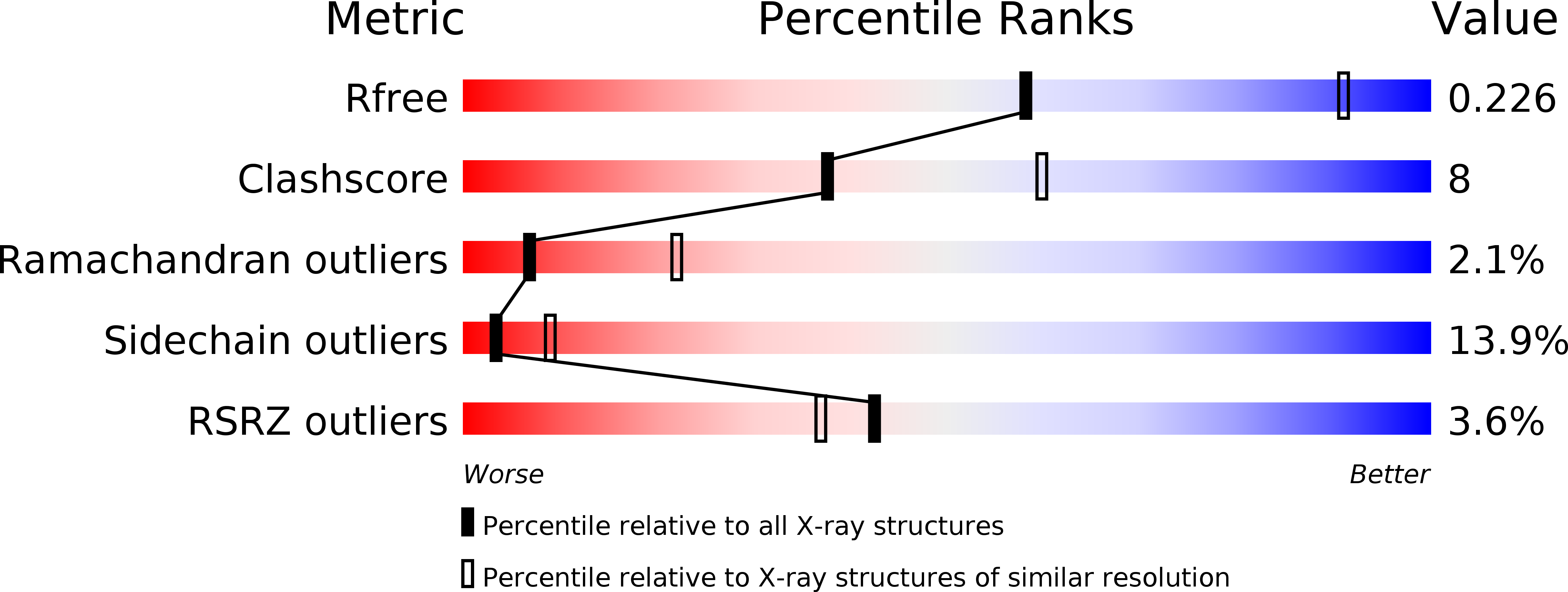
  • R-Value Free: 0.217 
  • R-Value Work: 0.161 
  • R-Value Observed: 0.164 

wwPDB Validation   3D Report Full Report


Ligand Structure Quality Assessment 


This is version 1.5 of the entry. See complete history


Literature

Molecular Blueprint of Allosteric Binding Sites in a Homologue of the Agonist-Binding Domain of the Alpha7 Nicotinic Acetylcholine Receptor.

Spurny, R.Debaveye, S.Farinha, A.Veys, K.Vos, A.M.Gossas, T.Atack, J.Bertrand, S.Bertrand, D.Danielson, U.H.Tresadern, G.Ulens, C.

(2015) Proc Natl Acad Sci U S A 112: E2543

  • DOI: https://doi.org/10.1073/pnas.1418289112
  • Primary Citation of Related Structures:  
    5AFH, 5AFJ, 5AFK, 5AFL, 5AFM, 5AFN

  • PubMed Abstract: 

    The α7 nicotinic acetylcholine receptor (nAChR) belongs to the family of pentameric ligand-gated ion channels and is involved in fast synaptic signaling. In this study, we take advantage of a recently identified chimera of the extracellular domain of the native α7 nicotinic acetylcholine receptor and acetylcholine binding protein, termed α7-AChBP. This chimeric receptor was used to conduct an innovative fragment-library screening in combination with X-ray crystallography to identify allosteric binding sites. One allosteric site is surface-exposed and is located near the N-terminal α-helix of the extracellular domain. Ligand binding at this site causes a conformational change of the α-helix as the fragment wedges between the α-helix and a loop homologous to the main immunogenic region of the muscle α1 subunit. A second site is located in the vestibule of the receptor, in a preexisting intrasubunit pocket opposite the agonist binding site and corresponds to a previously identified site involved in positive allosteric modulation of the bacterial homolog ELIC. A third site is located at a pocket right below the agonist binding site. Using electrophysiological recordings on the human α7 nAChR we demonstrate that the identified fragments, which bind at these sites, can modulate receptor activation. This work presents a structural framework for different allosteric binding sites in the α7 nAChR and paves the way for future development of novel allosteric modulators with therapeutic potential.


  • Organizational Affiliation

    Laboratory of Structural Neurobiology, Katholieke Universiteit Leuven, Leuven B-3000, Belgium;


Macromolecules
Find similar proteins by:  (by identity cutoff)  |  3D Structure
Entity ID: 1
MoleculeChains Sequence LengthOrganismDetailsImage
ACETYLCHOLINE-BINDING PROTEIN, NEURONAL ACETYLCHOLINE RECEPTOR SUBUNIT ALPHA-7
A, C, D, E
205Homo sapiensLymnaea stagnalis
This entity is chimeric
Mutation(s): 0 
UniProt & NIH Common Fund Data Resources
Find proteins for P58154 (Lymnaea stagnalis)
Explore P58154 
Go to UniProtKB:  P58154
Find proteins for P36544 (Homo sapiens)
Explore P36544 
Go to UniProtKB:  P36544
PHAROS:  P36544
GTEx:  ENSG00000175344 
Entity Groups  
Sequence Clusters30% Identity50% Identity70% Identity90% Identity95% Identity100% Identity
UniProt GroupsP36544P58154
Sequence Annotations
Expand
  • Reference Sequence
Find similar proteins by:  (by identity cutoff)  |  3D Structure
Entity ID: 2
MoleculeChains Sequence LengthOrganismDetailsImage
ACETYLCHOLINE-BINDING PROTEIN, NEURONAL ACETYLCHOLINE RECEPTOR SUBUNIT ALPHA-7207Homo sapiensLymnaea stagnalisMutation(s): 0 
UniProt & NIH Common Fund Data Resources
Find proteins for P58154 (Lymnaea stagnalis)
Explore P58154 
Go to UniProtKB:  P58154
Find proteins for P36544 (Homo sapiens)
Explore P36544 
Go to UniProtKB:  P36544
PHAROS:  P36544
GTEx:  ENSG00000175344 
Entity Groups  
Sequence Clusters30% Identity50% Identity70% Identity90% Identity95% Identity100% Identity
UniProt GroupsP36544P58154
Sequence Annotations
Expand
  • Reference Sequence
Small Molecules
Ligands 4 Unique
IDChains Name / Formula / InChI Key2D Diagram3D Interactions
L0B
Query on L0B

Download Ideal Coordinates CCD File 
AA [auth E],
H [auth A],
M [auth B],
Q [auth C],
V [auth D]
Alpha-Lobeline
C22 H27 N O2
MXYUKLILVYORSK-HBMCJLEFSA-N
9Z0
Query on 9Z0

Download Ideal Coordinates CCD File 
CA [auth E],
J [auth A],
N [auth B],
S [auth C],
X [auth D]
4,5-dibromo-N-(3-hydroxypropyl)-1H-pyrrole-2-carboxamide
C8 H10 Br2 N2 O2
UAVIBMSTKCBELE-UHFFFAOYSA-N
NAG
Query on NAG

Download Ideal Coordinates CCD File 
F [auth A]
G [auth A]
K [auth B]
L [auth B]
O [auth C]
F [auth A],
G [auth A],
K [auth B],
L [auth B],
O [auth C],
P [auth C],
T [auth D],
U [auth D],
Y [auth E],
Z [auth E]
2-acetamido-2-deoxy-beta-D-glucopyranose
C8 H15 N O6
OVRNDRQMDRJTHS-FMDGEEDCSA-N
GOL
Query on GOL

Download Ideal Coordinates CCD File 
BA [auth E],
I [auth A],
R [auth C],
W [auth D]
GLYCEROL
C3 H8 O3
PEDCQBHIVMGVHV-UHFFFAOYSA-N
Experimental Data & Validation

Experimental Data

  • Method: X-RAY DIFFRACTION
  • Resolution: 2.85 Å
  • R-Value Free: 0.217 
  • R-Value Work: 0.161 
  • R-Value Observed: 0.164 
  • Space Group: P 21 21 21
Unit Cell:
Length ( Å )Angle ( ˚ )
a = 75.7α = 90
b = 119.6β = 90
c = 143.3γ = 90
Software Package:
Software NamePurpose
BUSTERrefinement

Structure Validation

View Full Validation Report


Ligand Structure Quality Assessment 


Entry History 

Deposition Data

Revision History  (Full details and data files)

  • Version 1.0: 2015-05-06
    Type: Initial release
  • Version 1.1: 2015-05-13
    Changes: Database references
  • Version 1.2: 2015-05-27
    Changes: Database references
  • Version 1.3: 2017-03-15
    Changes: Source and taxonomy
  • Version 1.4: 2019-04-24
    Changes: Data collection, Source and taxonomy
  • Version 1.5: 2020-07-29
    Type: Remediation
    Reason: Carbohydrate remediation
    Changes: Data collection, Derived calculations, Other, Structure summary