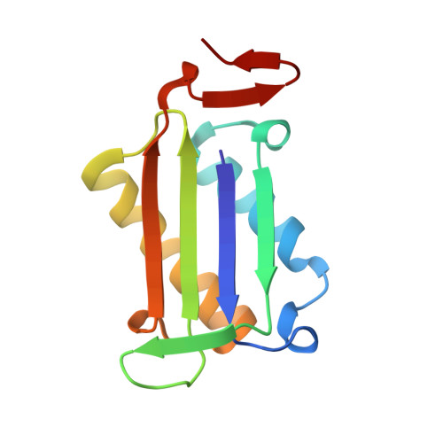
5BSI

Crystal structure of Y36A mutant of human macrophage migration inhibitory factor


Experimental Data Snapshot

  • Method: X-RAY DIFFRACTION
  • Resolution: 2.00 Å
  • R-Value Free: 0.221 
  • R-Value Work: 0.208 
  • R-Value Observed: 0.208 

Starting Model: experimental
View more details

wwPDB Validation   3D Report Full Report


This is version 1.1 of the entry. See complete history


Literature

An Analysis of MIF Structural Features that Control Functional Activation of CD74.

Pantouris, G.Syed, M.A.Fan, C.Rajasekaran, D.Cho, T.Y.Rosenberg, E.M.Bucala, R.Bhandari, V.Lolis, E.J.

(2015) Chem Biol 22: 1197-1205

  • DOI: https://doi.org/10.1016/j.chembiol.2015.08.006
  • Primary Citation of Related Structures:  
    4P01, 4P0H, 4PKZ, 4PLU, 4TRF, 4TRU, 4XX7, 4XX8, 5BS9, 5BSC, 5BSI

  • PubMed Abstract: 

    For more than 15 years, the tautomerase active site of macrophage migration inhibitory factor (MIF) and its catalytic residue Pro1 have been being targeted for the development of therapeutics that block activation of its cell surface receptor, CD74. Neither the biological role of the MIF catalytic site nor the mechanistic details of CD74 activation are well understood. The inherently unstable structure of CD74 remains the biggest obstacle in structural studies with MIF for understanding the basis of CD74 activation. Using a novel approach, we elucidate the mechanistic details that control activation of CD74 by MIF surface residues and identify structural parameters of inhibitors that reduce CD74 biological activation. We also find that N-terminal mutants located deep in the catalytic site affect surface residues immediately outside the catalytic site, which are responsible for reduction of CD74 activation.


  • Organizational Affiliation

    Department of Pharmacology, Yale School of Medicine, New Haven, CT 06510, USA.


Macromolecules
Find similar proteins by:  (by identity cutoff)  |  3D Structure
Entity ID: 1
MoleculeChains Sequence LengthOrganismDetailsImage
Macrophage migration inhibitory factor
A,
B,
C,
D,
E,
F,
G,
H [auth J]
114Homo sapiensMutation(s): 1 
Gene Names: MIFGLIFMMIF
EC: 5.3.2.1 (PDB Primary Data), 5.3.3.12 (PDB Primary Data)
UniProt & NIH Common Fund Data Resources
Find proteins for P14174 (Homo sapiens)
Explore P14174 
Go to UniProtKB:  P14174
PHAROS:  P14174
GTEx:  ENSG00000240972 
Entity Groups  
Sequence Clusters30% Identity50% Identity70% Identity90% Identity95% Identity100% Identity
UniProt GroupP14174
Sequence Annotations
Expand
  • Reference Sequence
Small Molecules
Ligands 2 Unique
IDChains Name / Formula / InChI Key2D Diagram3D Interactions
SO4
Query on SO4

Download Ideal Coordinates CCD File 
I [auth A]
K [auth B]
M [auth C]
O [auth D]
R [auth E]
I [auth A],
K [auth B],
M [auth C],
O [auth D],
R [auth E],
S [auth F],
U [auth G],
V [auth J]
SULFATE ION
O4 S
QAOWNCQODCNURD-UHFFFAOYSA-L
GOL
Query on GOL

Download Ideal Coordinates CCD File 
J [auth A]
L [auth B]
N [auth C]
P [auth D]
Q [auth D]
J [auth A],
L [auth B],
N [auth C],
P [auth D],
Q [auth D],
T [auth F]
GLYCEROL
C3 H8 O3
PEDCQBHIVMGVHV-UHFFFAOYSA-N
Experimental Data & Validation

Experimental Data

  • Method: X-RAY DIFFRACTION
  • Resolution: 2.00 Å
  • R-Value Free: 0.221 
  • R-Value Work: 0.208 
  • R-Value Observed: 0.208 
  • Space Group: H 3
Unit Cell:
Length ( Å )Angle ( ˚ )
a = 177.771α = 90
b = 177.771β = 90
c = 217.783γ = 120
Software Package:
Software NamePurpose
REFMACrefinement
HKL-2000data scaling
PDB_EXTRACTdata extraction
HKL-2000data reduction
PHASERphasing

Structure Validation

View Full Validation Report


Entry History 

Deposition Data

Revision History  (Full details and data files)

  • Version 1.0: 2015-09-30
    Type: Initial release
  • Version 1.1: 2023-09-27
    Changes: Data collection, Database references, Derived calculations, Refinement description