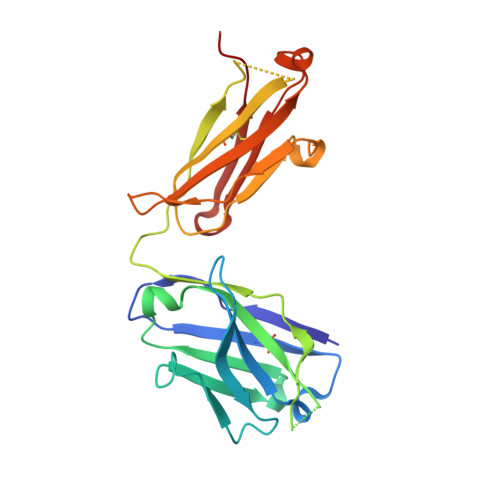
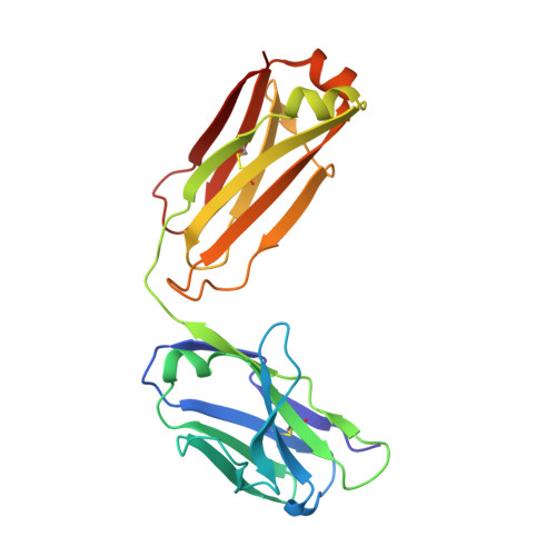
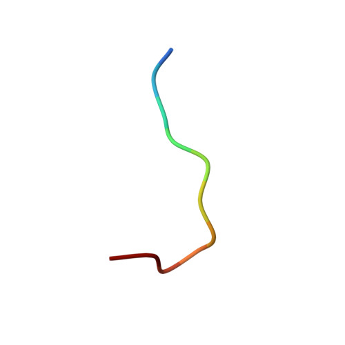
Gantenerumab: a novel human anti-Abeta antibody demonstrates sustained cerebral amyloid-Beta binding and elicits cell-mediated removal of human amyloid-Beta.
Bohrmann, B., Baumann, K., Benz, J., Gerber, F., Huber, W., Knoflach, F., Messer, J., Oroszlan, K., Rauchenberger, R., Richter, W.F., Rothe, C., Urban, M., Bardroff, M., Winter, M., Nordstedt, C., Loetscher, H.(2012) J Alzheimers Dis 28: 49-69
- PubMed: 21955818
- DOI: https://doi.org/10.3233/JAD-2011-110977
- Primary Citation of Related Structures:
5CSZ - PubMed Abstract:
The amyloid-β lowering capacity of anti-Aβ antibodies has been demonstrated in transgenic models of Alzheimer's disease (AD) and in AD patients. While the mechanism of immunotherapeutic amyloid-β removal is controversial, antibody-mediated sequestration of peripheral Aβ versus microglial phagocytic activity and disassembly of cerebral amyloid (or a combination thereof) has been proposed. For successful Aβ immunotherapy, we hypothesized that high affinity antibody binding to amyloid-β plaques and recruitment of brain effector cells is required for most efficient amyloid clearance. Here we report the generation of a novel fully human anti-Aβ antibody, gantenerumab, optimized in vitro for binding with sub-nanomolar affinity to a conformational epitope expressed on amyloid-β fibrils using HuCAL(®) phage display technologies. In peptide maps, both N-terminal and central portions of Aβ were recognized by gantenerumab. Remarkably, a novel orientation of N-terminal Aβ bound to the complementarity determining regions was identified by x-ray analysis of a gantenerumab Fab-Aβ(1-11) complex. In functional assays gantenerumab induced cellular phagocytosis of human amyloid-β deposits in AD brain slices when co-cultured with primary human macrophages and neutralized oligomeric Aβ42-mediated inhibitory effects on long-term potentiation in rat brain. In APP751(swedish)xPS2(N141I) transgenic mice, gantenerumab showed sustained binding to cerebral amyloid-β and, upon chronic treatment, significantly reduced small amyloid-β plaques by recruiting microglia and prevented new plaque formation. Unlike other Aβ antibodies, gantenerumab did not alter plasma Aβ suggesting undisturbed systemic clearance of soluble Aβ. These studies demonstrated that gantenerumab preferentially interacts with aggregated Aβ in the brain and lowers amyloid-β by eliciting effector cell-mediated clearance.
Organizational Affiliation:
F. Hoffmann-La Roche Ltd., pRED, Basel, Switzerland. bernd.bohrmann@roche.com


