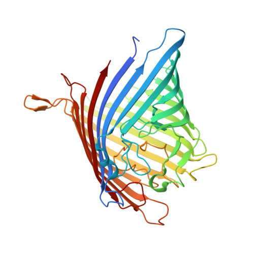
5DL7

Crystal structure of Acinetobacter baumannii OccAB3


Experimental Data Snapshot

  • Method: X-RAY DIFFRACTION
  • Resolution: 1.75 Å
  • R-Value Free: 0.191 
  • R-Value Work: 0.168 
  • R-Value Observed: 0.169 

Starting Model: experimental
View more details

wwPDB Validation   3D Report Full Report


Ligand Structure Quality Assessment 


This is version 1.2 of the entry. See complete history


Literature

Structural Insights into Outer Membrane Permeability of Acinetobacter baumannii.

Zahn, M.Bhamidimarri, S.P.Basle, A.Winterhalter, M.van den Berg, B.

(2016) Structure 24: 221-231

  • DOI: https://doi.org/10.1016/j.str.2015.12.009
  • Primary Citation of Related Structures:  
    5DL5, 5DL6, 5DL7, 5DL8

  • PubMed Abstract: 

    Bacterial resistance against antibiotics is an increasing global health problem. In Gram-negative bacteria the low permeability of the outer membrane (OM) is a major factor contributing to resistance, making it important to understand channel-mediated small-molecule passage of the OM. Acinetobacter baumannii has five Occ (OM carboxylate channel) proteins, which collectively are of major importance for the entry of small molecules. To improve our understanding of the OM permeability of A. baumannii, we present here the X-ray crystal structures of four Occ proteins, renamed OccAB1 to OccAB4. In addition we have carried out a biochemical and biophysical characterization using electrophysiology and liposome swelling experiments, providing information on substrate specificities. We identify OccAB1 as having the largest pore of the Occ proteins with corresponding high rates of small-molecule uptake, and we suggest that the future design of efficient antibiotics should focus on scaffolds that can permeate efficiently through the OccAB1 channel.


  • Organizational Affiliation

    Institute for Cellular and Molecular Biosciences, Medical School, Newcastle University, Newcastle upon Tyne NE2 4HH, UK.


Macromolecules
Find similar proteins by:  (by identity cutoff)  |  3D Structure
Entity ID: 1
MoleculeChains Sequence LengthOrganismDetailsImage
Porin419Acinetobacter baumannii AB307-0294Mutation(s): 0 
Gene Names: ABUW_2771
Membrane Entity: Yes 
UniProt
Find proteins for A0A0D5YKR4 (Acinetobacter baumannii)
Explore A0A0D5YKR4 
Go to UniProtKB:  A0A0D5YKR4
Entity Groups  
Sequence Clusters30% Identity50% Identity70% Identity90% Identity95% Identity100% Identity
UniProt GroupA0A0D5YKR4
Sequence Annotations
Expand
  • Reference Sequence
Small Molecules
Ligands 2 Unique
IDChains Name / Formula / InChI Key2D Diagram3D Interactions
C8E
Query on C8E

Download Ideal Coordinates CCD File 
F [auth A]
G [auth A]
H [auth A]
I [auth A]
J [auth A]
F [auth A],
G [auth A],
H [auth A],
I [auth A],
J [auth A],
K [auth A],
L [auth A]
(HYDROXYETHYLOXY)TRI(ETHYLOXY)OCTANE
C16 H34 O5
FEOZZFHAVXYAMB-UHFFFAOYSA-N
CA
Query on CA

Download Ideal Coordinates CCD File 
B [auth A],
C [auth A],
D [auth A],
E [auth A]
CALCIUM ION
Ca
BHPQYMZQTOCNFJ-UHFFFAOYSA-N
Experimental Data & Validation

Experimental Data

  • Method: X-RAY DIFFRACTION
  • Resolution: 1.75 Å
  • R-Value Free: 0.191 
  • R-Value Work: 0.168 
  • R-Value Observed: 0.169 
  • Space Group: I 2 2 2
Unit Cell:
Length ( Å )Angle ( ˚ )
a = 81.563α = 90
b = 125.717β = 90
c = 137.459γ = 90
Software Package:
Software NamePurpose
REFMACrefinement
XDSdata reduction
Aimlessdata scaling
MOLREPphasing

Structure Validation

View Full Validation Report


Ligand Structure Quality Assessment 


Entry History 

Deposition Data

Revision History  (Full details and data files)

  • Version 1.0: 2016-02-03
    Type: Initial release
  • Version 1.1: 2016-02-10
    Changes: Database references
  • Version 1.2: 2024-01-10
    Changes: Data collection, Database references, Derived calculations, Refinement description