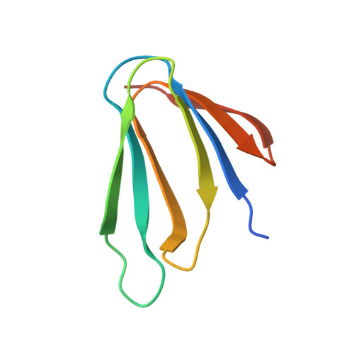
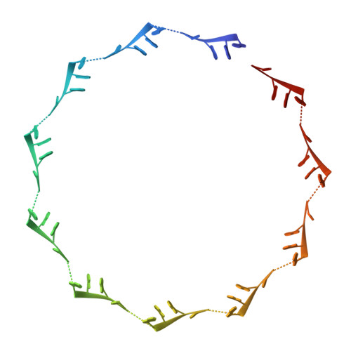
5EEW

RADIATION DAMAGE TO THE TRAP-RNA COMPLEX: DOSE (DWD) 6.45 MGy


Experimental Data Snapshot

  • Method: X-RAY DIFFRACTION
  • Resolution: 1.98 Å
  • R-Value Free: 0.243 
  • R-Value Work: 0.209 
  • R-Value Observed: 0.211 

Starting Model: experimental
View more details

wwPDB Validation   3D Report Full Report


Ligand Structure Quality Assessment 


This is version 1.3 of the entry. See complete history


Literature

RNA protects a nucleoprotein complex against radiation damage.

Bury, C.S.McGeehan, J.E.Antson, A.A.Carmichael, I.Gerstel, M.Shevtsov, M.B.Garman, E.F.

(2016) Acta Crystallogr D Struct Biol 72: 648-657

  • DOI: https://doi.org/10.1107/S2059798316003351
  • Primary Citation of Related Structures:  
    5EEU, 5EEV, 5EEW, 5EEX, 5EEY, 5EEZ, 5EF0, 5EF1, 5EF2, 5EF3

  • PubMed Abstract: 

    Radiation damage during macromolecular X-ray crystallographic data collection is still the main impediment for many macromolecular structure determinations. Even when an eventual model results from the crystallographic pipeline, the manifestations of radiation-induced structural and conformation changes, the so-called specific damage, within crystalline macromolecules can lead to false interpretations of biological mechanisms. Although this has been well characterized within protein crystals, far less is known about specific damage effects within the larger class of nucleoprotein complexes. Here, a methodology has been developed whereby per-atom density changes could be quantified with increasing dose over a wide (1.3-25.0 MGy) range and at higher resolution (1.98 Å) than the previous systematic specific damage study on a protein-DNA complex. Specific damage manifestations were determined within the large trp RNA-binding attenuation protein (TRAP) bound to a single-stranded RNA that forms a belt around the protein. Over a large dose range, the RNA was found to be far less susceptible to radiation-induced chemical changes than the protein. The availability of two TRAP molecules in the asymmetric unit, of which only one contained bound RNA, allowed a controlled investigation into the exact role of RNA binding in protein specific damage susceptibility. The 11-fold symmetry within each TRAP ring permitted statistically significant analysis of the Glu and Asp damage patterns, with RNA binding unexpectedly being observed to protect these otherwise highly sensitive residues within the 11 RNA-binding pockets distributed around the outside of the protein molecule. Additionally, the method enabled a quantification of the reduction in radiation-induced Lys and Phe disordering upon RNA binding directly from the electron density.


  • Organizational Affiliation

    Laboratory of Molecular Biophysics, Department of Biochemistry, University of Oxford, South Parks Road, Oxford OX1 3QU, England.


Macromolecules

Find similar proteins by:  (by identity cutoff)  |  3D Structure
Entity ID: 1
MoleculeChains Sequence LengthOrganismDetailsImage
Transcription attenuation protein MtrB
A, B, C, D, E
A, B, C, D, E, F, G, H, I, J, K, L, M, N, O, P, Q, R, S, T, U, V
74Geobacillus stearothermophilusMutation(s): 0 
Gene Names: mtrB
UniProt
Find proteins for Q9X6J6 (Geobacillus stearothermophilus)
Explore Q9X6J6 
Go to UniProtKB:  Q9X6J6
Entity Groups  
Sequence Clusters30% Identity50% Identity70% Identity90% Identity95% Identity100% Identity
UniProt GroupQ9X6J6
Sequence Annotations
Expand
  • Reference Sequence
Find similar nucleic acids by:  (by identity cutoff)  |  3D Structure
Entity ID: 2
MoleculeChains LengthOrganismImage
(GAGUU)10GAG 53-NUCLEOTIDE RNA55synthetic construct
Sequence Annotations
Expand
  • Reference Sequence
Small Molecules
Ligands 1 Unique
IDChains Name / Formula / InChI Key2D Diagram3D Interactions
TRP
Query on TRP

Download Ideal Coordinates CCD File 
AA [auth D]
BA [auth E]
CA [auth F]
DA [auth G]
EA [auth H]
AA [auth D],
BA [auth E],
CA [auth F],
DA [auth G],
EA [auth H],
FA [auth I],
GA [auth J],
HA [auth K],
IA [auth L],
JA [auth M],
KA [auth N],
LA [auth O],
MA [auth P],
NA [auth Q],
OA [auth R],
PA [auth S],
QA [auth T],
RA [auth U],
SA [auth V],
X [auth A],
Y [auth B],
Z [auth C]
TRYPTOPHAN
C11 H12 N2 O2
QIVBCDIJIAJPQS-VIFPVBQESA-N
Experimental Data & Validation

Experimental Data

  • Method: X-RAY DIFFRACTION
  • Resolution: 1.98 Å
  • R-Value Free: 0.243 
  • R-Value Work: 0.209 
  • R-Value Observed: 0.211 
  • Space Group: C 1 2 1
Unit Cell:
Length ( Å )Angle ( ˚ )
a = 141.04α = 90
b = 111.02β = 117.41
c = 137.99γ = 90
Software Package:
Software NamePurpose
MOSFLMdata reduction
Aimlessdata scaling
PHENIXrefinement
PDB_EXTRACTdata extraction
PHASERphasing

Structure Validation

View Full Validation Report


Ligand Structure Quality Assessment 


Entry History & Funding Information

Deposition Data


Funding OrganizationLocationGrant Number
Engineering and Physical Sciences Research CouncilUnited Kingdom--

Revision History  (Full details and data files)

  • Version 1.0: 2016-05-04
    Type: Initial release
  • Version 1.1: 2017-09-13
    Changes: Author supporting evidence, Data collection
  • Version 1.2: 2017-11-22
    Changes: Database references
  • Version 1.3: 2024-01-10
    Changes: Data collection, Database references, Refinement description