5HWR

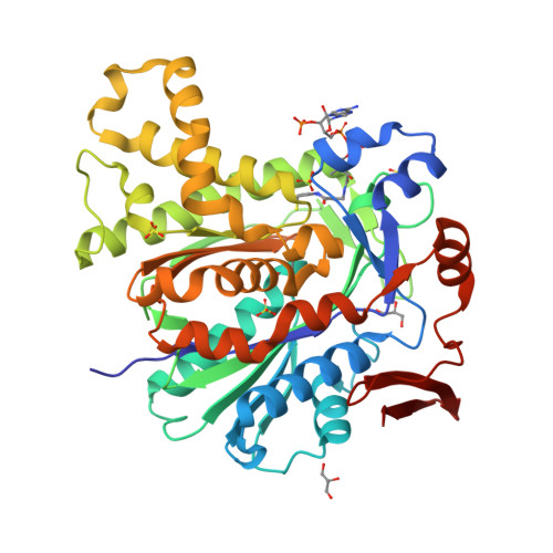
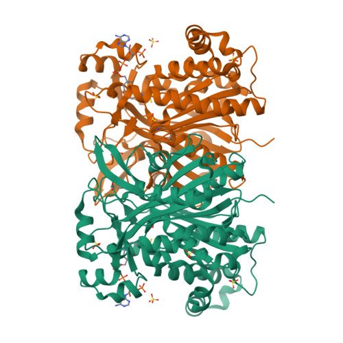
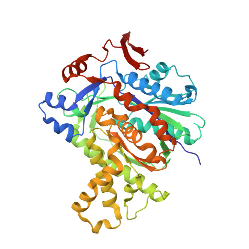
MvaS in complex with coenzyme A


Experimental Data Snapshot

  • Method: X-RAY DIFFRACTION
  • Resolution: 1.50 Å
  • R-Value Free: 0.157 
  • R-Value Work: 0.124 
  • R-Value Observed: 0.126 

wwPDB Validation   3D Report Full Report


Ligand Structure Quality Assessment 


This is version 1.2 of the entry. See complete history


Literature

Crystal Structure of the HMG-CoA Synthase MvaS from the Gram-Negative Bacterium Myxococcus xanthus.

Bock, T.Kasten, J.Muller, R.Blankenfeldt, W.

(2016) Chembiochem 17: 1257-1262

  • DOI: https://doi.org/10.1002/cbic.201600070
  • Primary Citation of Related Structures:  
    5HWO, 5HWP, 5HWQ, 5HWR

  • PubMed Abstract: 

    A critical step in bacterial isoprenoid production is the synthesis of 3-hydroxy-3-methylglutaryl coenzyme A (HMG-CoA) catalyzed by HMG-CoA synthase (HMGCS). In myxobacteria, this enzyme is also involved in a recently discovered alternative and acetyl-CoA-dependent isovaleryl CoA biosynthesis pathway. Here we present crystal structures of MvaS, the HMGCS from Myxococcus xanthus, in complex with CoA and acetylated active site Cys115, with the second substrate acetoacetyl CoA and with the product of the condensation reaction, 3-hydroxy-3-methylglutaryl CoA. With these structures, we show that MvaS uses the common HMGCS enzymatic mechanism and provide evidence that dimerization plays a role in the formation and stability of the active site. Overall, MvaS shows features typical of the eukaryotic HMGCS and exhibits differences from homologues from Gram-positive bacteria. This study provides insights into myxobacterial alternative isovaleryl CoA biosynthesis and thereby extends the toolbox for the biotechnological production of renewable fuel and chemicals.


  • Organizational Affiliation

    Structure and Function of Proteins, Helmholtz Centre for Infection Research, Inhoffenstrasse 7, 38124, Braunschweig, Germany.


Macromolecules
Find similar proteins by:  (by identity cutoff)  |  3D Structure
Entity ID: 1
MoleculeChains Sequence LengthOrganismDetailsImage
Hydroxymethylglutaryl-CoA synthase420Myxococcus xanthus DK 1622Mutation(s): 0 
Gene Names: mvaSMXAN_4267
EC: 2.3.3.10
UniProt
Find proteins for Q1D4I1 (Myxococcus xanthus (strain DK1622))
Explore Q1D4I1 
Go to UniProtKB:  Q1D4I1
Entity Groups  
Sequence Clusters30% Identity50% Identity70% Identity90% Identity95% Identity100% Identity
UniProt GroupQ1D4I1
Sequence Annotations
Expand
  • Reference Sequence
Small Molecules
Ligands 3 Unique
IDChains Name / Formula / InChI Key2D Diagram3D Interactions
COA
Query on COA

Download Ideal Coordinates CCD File 
H [auth A]COENZYME A
C21 H36 N7 O16 P3 S
RGJOEKWQDUBAIZ-IBOSZNHHSA-N
SO4
Query on SO4

Download Ideal Coordinates CCD File 
D [auth A],
E [auth A],
F [auth A],
G [auth A]
SULFATE ION
O4 S
QAOWNCQODCNURD-UHFFFAOYSA-L
GOL
Query on GOL

Download Ideal Coordinates CCD File 
B [auth A],
C [auth A]
GLYCEROL
C3 H8 O3
PEDCQBHIVMGVHV-UHFFFAOYSA-N
Modified Residues  1 Unique
IDChains TypeFormula2D DiagramParent
CSD
Query on CSD
A
L-PEPTIDE LINKINGC3 H7 N O4 SCYS
Experimental Data & Validation

Experimental Data

  • Method: X-RAY DIFFRACTION
  • Resolution: 1.50 Å
  • R-Value Free: 0.157 
  • R-Value Work: 0.124 
  • R-Value Observed: 0.126 
  • Space Group: P 61 2 2
Unit Cell:
Length ( Å )Angle ( ˚ )
a = 73.162α = 90
b = 73.162β = 90
c = 276.899γ = 120
Software Package:
Software NamePurpose
PHENIXrefinement
Aimlessdata scaling
PDB_EXTRACTdata extraction
XDSdata reduction
PHENIXphasing

Structure Validation

View Full Validation Report


Ligand Structure Quality Assessment 


Entry History 

Deposition Data

Revision History  (Full details and data files)

  • Version 1.0: 2016-05-11
    Type: Initial release
  • Version 1.1: 2016-07-13
    Changes: Database references
  • Version 1.2: 2024-10-16
    Changes: Data collection, Database references, Structure summary