5NC9

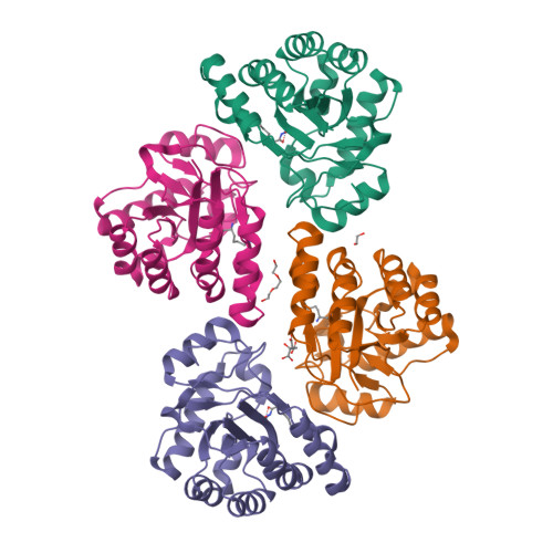
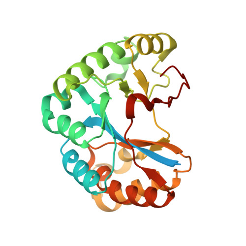
Crystal structure of the polysaccharide deacetylase Bc1974 from Bacillus cereus in complex with (2S)-2,6-diamino-N-hydroxyhexanamide


Experimental Data Snapshot

  • Method: X-RAY DIFFRACTION
  • Resolution: 2.44 Å
  • R-Value Free: 0.279 
  • R-Value Work: 0.220 
  • R-Value Observed: 0.223 

wwPDB Validation   3D Report Full Report


Ligand Structure Quality Assessment 


This is version 1.1 of the entry. See complete history


Literature

Structures of the Peptidoglycan N-Acetylglucosamine Deacetylase Bc1974 and Its Complexes with Zinc Metalloenzyme Inhibitors.

Giastas, P.Andreou, A.Papakyriakou, A.Koutsioulis, D.Balomenou, S.Tzartos, S.J.Bouriotis, V.Eliopoulos, E.E.

(2018) Biochemistry 57: 753-763

  • DOI: https://doi.org/10.1021/acs.biochem.7b00919
  • Primary Citation of Related Structures:  
    5N1J, 5N1P, 5NC6, 5NC9, 5NCD, 5NEK, 5NEL

  • PubMed Abstract: 

    The cell wall peptidoglycan is recognized as a primary target of the innate immune system, and usually its disintegration results in bacterial lysis. Bacillus cereus, a close relative of the highly virulent Bacillus anthracis, contains 10 polysaccharide deacetylases. Among these, the peptidoglycan N-acetylglucosamine deacetylase Bc1974 is the highest homologue to the Bacillus anthracis Ba1977 that is required for full virulence and is involved in resistance to the host's lysozyme. These metalloenzymes belong to the carbohydrate esterase family 4 (CE4) and are attractive targets for the development of new anti-infective agents. Herein we report the first X-ray crystal structures of the NodB domain of Bc1974, the conserved catalytic core of CE4s, in the unliganded form and in complex with four known metalloenzyme inhibitors and two amino acid hydroxamates that target the active site metal. These structures revealed the presence of two conformational states of a catalytic loop known as motif-4 (MT4), which were not observed previously for peptidoglycan deacetylases, but were recently shown in the structure of a Vibrio clolerae chitin deacetylase. By employing molecular docking of a substrate model, we describe a catalytic mechanism that probably involves initial binding of the substrate in a receptive, more open state of MT4 and optimal catalytic activity in the closed state of MT4, consistent with the previous observations. The ligand-bound structures presented here, in addition to the five Bc1974 inhibitors identified, provide a valuable basis for the design of antibacterial agents that target the peptidoglycan deacetylase Ba1977.


  • Organizational Affiliation

    Department of Biotechnology, Laboratory of Genetics, Agricultural University of Athens , Iera Odos 75, 11855 Athens, Greece.


Macromolecules
Find similar proteins by:  (by identity cutoff)  |  3D Structure
Entity ID: 1
MoleculeChains Sequence LengthOrganismDetailsImage
Peptidoglycan N-acetylglucosamine deacetylase
A, B, C, D
247Bacillus cereus ATCC 14579Mutation(s): 0 
Gene Names: BC_1974
EC: 3.5.1 (PDB Primary Data), 3.5.1.104 (UniProt)
UniProt
Find proteins for Q81EJ6 (Bacillus cereus (strain ATCC 14579 / DSM 31 / CCUG 7414 / JCM 2152 / NBRC 15305 / NCIMB 9373 / NCTC 2599 / NRRL B-3711))
Explore Q81EJ6 
Go to UniProtKB:  Q81EJ6
Entity Groups  
Sequence Clusters30% Identity50% Identity70% Identity90% Identity95% Identity100% Identity
UniProt GroupQ81EJ6
Sequence Annotations
Expand
  • Reference Sequence
Small Molecules
Ligands 5 Unique
IDChains Name / Formula / InChI Key2D Diagram3D Interactions
CIT
Query on CIT

Download Ideal Coordinates CCD File 
I [auth B]CITRIC ACID
C6 H8 O7
KRKNYBCHXYNGOX-UHFFFAOYSA-N
8SZ
Query on 8SZ

Download Ideal Coordinates CCD File 
E [auth A],
G [auth B],
K [auth C],
M [auth D]
(2~{S})-2,6-bis(azanyl)-~{N}-oxidanyl-hexanamide
C6 H15 N3 O2
NZWPVDFOIUKVSJ-YFKPBYRVSA-N
PGE
Query on PGE

Download Ideal Coordinates CCD File 
O [auth D]TRIETHYLENE GLYCOL
C6 H14 O4
ZIBGPFATKBEMQZ-UHFFFAOYSA-N
ZN
Query on ZN

Download Ideal Coordinates CCD File 
F [auth A],
H [auth B],
L [auth C],
N [auth D]
ZINC ION
Zn
PTFCDOFLOPIGGS-UHFFFAOYSA-N
EDO
Query on EDO

Download Ideal Coordinates CCD File 
J [auth B]1,2-ETHANEDIOL
C2 H6 O2
LYCAIKOWRPUZTN-UHFFFAOYSA-N
Experimental Data & Validation

Experimental Data

  • Method: X-RAY DIFFRACTION
  • Resolution: 2.44 Å
  • R-Value Free: 0.279 
  • R-Value Work: 0.220 
  • R-Value Observed: 0.223 
  • Space Group: P 1 21 1
Unit Cell:
Length ( Å )Angle ( ˚ )
a = 49.767α = 90
b = 117.835β = 102.38
c = 99.059γ = 90
Software Package:
Software NamePurpose
PHENIXrefinement
XDSdata reduction
XDSdata scaling
PHASERphasing

Structure Validation

View Full Validation Report


Ligand Structure Quality Assessment 


Entry History 

Deposition Data

Revision History  (Full details and data files)

  • Version 1.0: 2018-02-21
    Type: Initial release
  • Version 1.1: 2024-05-08
    Changes: Data collection, Database references, Derived calculations