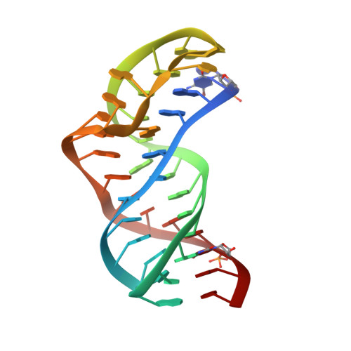
5NZD

The structure of the thermobifida fusca guanidine III riboswitch in space group P212121.


Experimental Data Snapshot

  • Method: X-RAY DIFFRACTION
  • Resolution: 2.01 Å
  • R-Value Free: 0.245 
  • R-Value Work: 0.220 
  • R-Value Observed: 0.221 

wwPDB Validation   3D Report Full Report


This is version 1.2 of the entry. See complete history


Literature

Structure of the Guanidine III Riboswitch.

Huang, L.Wang, J.Wilson, T.J.Lilley, D.M.J.

(2017) Cell Chem Biol 24: 1407-1415.e2

  • DOI: https://doi.org/10.1016/j.chembiol.2017.08.021
  • Primary Citation of Related Structures:  
    5NWQ, 5NY8, 5NZ3, 5NZ6, 5NZD, 5O62, 5O69

  • PubMed Abstract: 

    Riboswitches are structural elements found in mRNA molecules that couple small-molecule binding to regulation of gene expression, usually by controlling transcription or translation. We have determined high-resolution crystal structures of the ykkC guanidine III riboswitch from Thermobifida fusca. The riboswitch forms a classic H-type pseudoknot that includes a triple helix that is continuous with a central core of conserved nucleotides. These form a left-handed helical ramp of inter-nucleotide interactions, generating the guanidinium cation binding site. The ligand is hydrogen bonded to the Hoogsteen edges of two guanine bases. The binding pocket has a side opening that can accommodate a small side chain, shown by structures with bound methylguanidine, aminoguanidine, ethylguanidine, and agmatine. Comparison of the new structure with those of the guanidine I and II riboswitches reveals that evolution generated three different structural solutions for guanidine binding and subsequent gene regulation, although with some common elements.


  • Organizational Affiliation

    Cancer Research UK Nucleic Acid Structure Research Group, MSI/WTB Complex, The University of Dundee, Dow Street, Dundee DD1 5EH, UK.


Macromolecules
Find similar nucleic acids by:  (by identity cutoff)  |  3D Structure
Entity ID: 1
MoleculeChains LengthOrganismImage
guanidine III riboswitch
A, B
41Thermobifida fusca
Sequence Annotations
Expand
  • Reference Sequence
Small Molecules
Ligands 3 Unique
IDChains Name / Formula / InChI Key2D Diagram3D Interactions
ACT
Query on ACT

Download Ideal Coordinates CCD File 
J [auth A],
O [auth B]
ACETATE ION
C2 H3 O2
QTBSBXVTEAMEQO-UHFFFAOYSA-M
MG
Query on MG

Download Ideal Coordinates CCD File 
C [auth A]
D [auth A]
E [auth A]
F [auth A]
G [auth A]
C [auth A],
D [auth A],
E [auth A],
F [auth A],
G [auth A],
K [auth B],
L [auth B],
M [auth B],
N [auth B]
MAGNESIUM ION
Mg
JLVVSXFLKOJNIY-UHFFFAOYSA-N
NA
Query on NA

Download Ideal Coordinates CCD File 
H [auth A],
I [auth A]
SODIUM ION
Na
FKNQFGJONOIPTF-UHFFFAOYSA-N
Experimental Data & Validation

Experimental Data

  • Method: X-RAY DIFFRACTION
  • Resolution: 2.01 Å
  • R-Value Free: 0.245 
  • R-Value Work: 0.220 
  • R-Value Observed: 0.221 
  • Space Group: P 21 21 21
Unit Cell:
Length ( Å )Angle ( ˚ )
a = 49.46α = 90
b = 64.613β = 90
c = 84.292γ = 90
Software Package:
Software NamePurpose
PHENIXrefinement
xia2data reduction
xia2data scaling
PHENIXphasing

Structure Validation

View Full Validation Report


Entry History & Funding Information

Deposition Data


Funding OrganizationLocationGrant Number
Cancer Research UKUnited Kingdomprogram grant A18604

Revision History  (Full details and data files)

  • Version 1.0: 2017-10-18
    Type: Initial release
  • Version 1.1: 2017-11-29
    Changes: Database references
  • Version 1.2: 2024-05-08
    Changes: Data collection, Database references, Derived calculations