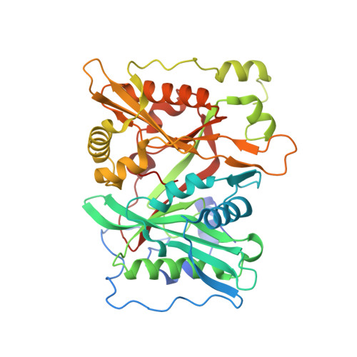
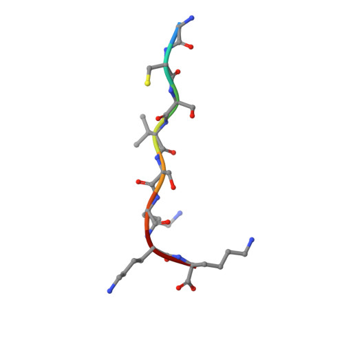
5O9U

HsNMT1 in complex with CoA and Myristoylated-GCSVSKKK octapeptide


Experimental Data Snapshot

  • Method: X-RAY DIFFRACTION
  • Resolution: 1.85 Å
  • R-Value Free: 0.192 
  • R-Value Work: 0.155 
  • R-Value Observed: 0.157 

Starting Model: experimental
View more details

wwPDB Validation   3D Report Full Report


Ligand Structure Quality Assessment 


This is version 1.3 of the entry. See complete history


Literature

Structural and genomic decoding of human and plant myristoylomes reveals a definitive recognition pattern.

Castrec, B.Dian, C.Ciccone, S.Ebert, C.L.Bienvenut, W.V.Le Caer, J.P.Steyaert, J.M.Giglione, C.Meinnel, T.

(2018) Nat Chem Biol 14: 671-679

  • DOI: https://doi.org/10.1038/s41589-018-0077-5
  • Primary Citation of Related Structures:  
    5O9S, 5O9T, 5O9U, 5O9V

  • PubMed Abstract: 

    An organism's entire protein modification repertoire has yet to be comprehensively mapped. N-myristoylation (MYR) is a crucial eukaryotic N-terminal protein modification. Here we mapped complete Homo sapiens and Arabidopsis thaliana myristoylomes. The crystal structures of human modifier NMT1 complexed with reactive and nonreactive target-mimicking peptide ligands revealed unexpected binding clefts and a modifier recognition pattern. This information allowed integrated mapping of myristoylomes using peptide macroarrays, dedicated prediction algorithms, and in vivo mass spectrometry. Global MYR profiling at the genomic scale identified over a thousand novel, heterogeneous targets in both organisms. Surprisingly, MYR involved a non-negligible set of overlapping targets with N-acetylation, and the sequence signature marks for a third proximal acylation-S-palmitoylation-were genomically imprinted, allowing recognition of sequences exhibiting both acylations. Together, the data extend the N-end rule concept for Gly-starting proteins to subcellular compartmentalization and reveal the main neighbors influencing protein modification profiles and consequent cell fate.


  • Organizational Affiliation

    Institute for Integrative Biology of the Cell (I2BC), CEA, CNRS, Univ. Paris-Sud, Université Paris Saclay, Gif-sur-Yvette cedex, France.


Macromolecules
Find similar proteins by:  (by identity cutoff)  |  3D Structure
Entity ID: 1
MoleculeChains Sequence LengthOrganismDetailsImage
Glycylpeptide N-tetradecanoyltransferase 1
A, B
402Homo sapiensMutation(s): 0 
Gene Names: NMT1NMT
EC: 2.3.1.97
UniProt & NIH Common Fund Data Resources
Find proteins for P30419 (Homo sapiens)
Go to UniProtKB:  P30419
PHAROS:  P30419
GTEx:  ENSG00000136448 
Sequence Annotations
Expand
  • Reference Sequence

Find similar proteins by:  Sequence   |   3D Structure  

Entity ID: 2
MoleculeChains Sequence LengthOrganismDetailsImage
Calcineurin B-like protein 4
C, D
8Arabidopsis thalianaMutation(s): 0 
UniProt
Find proteins for O81223 (Arabidopsis thaliana)
Go to UniProtKB:  O81223
Sequence Annotations
Expand
  • Reference Sequence
Experimental Data & Validation

Experimental Data

  • Method: X-RAY DIFFRACTION
  • Resolution: 1.85 Å
  • R-Value Free: 0.192 
  • R-Value Work: 0.155 
  • R-Value Observed: 0.157 
  • Space Group: P 2 21 21
Unit Cell:
Length ( Å )Angle ( ˚ )
a = 58.236α = 90
b = 79.779β = 90
c = 178.751γ = 90
Software Package:
Software NamePurpose
PHENIXrefinement
XDSdata reduction
Aimlessdata scaling
MOLREPphasing

Structure Validation

View Full Validation Report


Ligand Structure Quality Assessment 


Entry History & Funding Information

Deposition Data


Funding OrganizationLocationGrant Number
French National Research AgencyFranceANR PalmyProt
Fondation ARCFranceSFI2011120111203841

Revision History  (Full details and data files)

  • Version 1.0: 2018-06-27
    Type: Initial release
  • Version 1.1: 2018-11-07
    Changes: Data collection, Database references
  • Version 1.2: 2024-01-17
    Changes: Data collection, Database references, Refinement description
  • Version 1.3: 2024-10-23
    Changes: Structure summary