5T8U | pdb_00005t8u

Crystal structure of P. falciparum LipL1 in complex lipoate


Experimental Data Snapshot

  • Method: X-RAY DIFFRACTION
  • Resolution: 2.32 Å
  • R-Value Free: 
    0.302 (Depositor), 0.300 (DCC) 
  • R-Value Work: 
    0.273 (Depositor), 0.270 (DCC) 
  • R-Value Observed: 
    0.274 (Depositor) 

Starting Model: experimental
View more details

wwPDB Validation   3D Report Full Report


Ligand Structure Quality Assessment 

Created with Raphaël 2.3.0Worse 01 BetterLigand structure goodness of fit to experimental dataBest fitted LPAClick on this verticalbar to view details

This is version 1.5 of the entry. See complete history


Literature

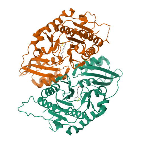
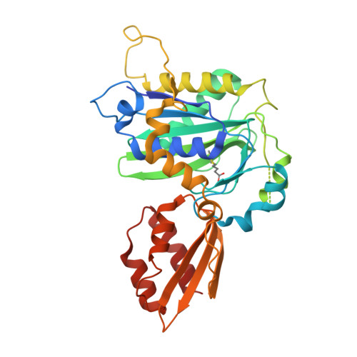
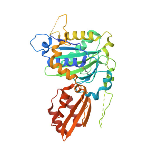
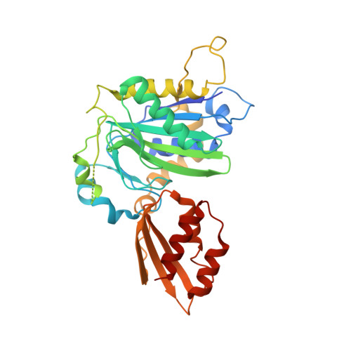
Crystal structure of lipoate-bound lipoate ligase 1, LipL1, from Plasmodium falciparum.

Guerra, A.J.Afanador, G.A.Prigge, S.T.

(2017) Proteins 85: 1777-1783

  • DOI: https://doi.org/10.1002/prot.25324
  • Primary Citation of Related Structures:  
    5T8U

  • PubMed Abstract: 

    Plasmodium falciparum lipoate protein ligase 1 (PfLipL1) is an ATP-dependent ligase that belongs to the biotin/lipoate A/B protein ligase family (PFAM PF03099). PfLipL1 is the only known canonical lipoate ligase in Pf and functions as a redox switch between two lipoylation routes in the parasite mitochondrion. Here, we report the crystal structure of a deletion construct of PfLipL1 (PfLipL1 Δ243-279 ) bound to lipoate, and validate the lipoylation activity of this construct in both an in vitro lipoylation assay and a cell-based lipoylation assay. This characterization represents the first step in understanding the redox dependence of the lipoylation mechanism in malaria parasites. Proteins 2017; 85:1777-1783. © 2017 Wiley Periodicals, Inc.


  • Organizational Affiliation

    Department of Molecular Microbiology and Immunology, Johns Hopkins Bloomberg School of Public Health, Baltimore, Maryland.


Macromolecules
Find similar proteins by:  (by identity cutoff)  |  3D Structure
Entity ID: 1
MoleculeChains Sequence LengthOrganismDetailsImage
Lipoate-protein ligase 1
A, B
360Plasmodium falciparum 3D7Mutation(s): 0 
Gene Names: PF3D7_1314600
EC: 2.7.7.63 (PDB Primary Data), 6.3.1.20 (UniProt)
UniProt
Find proteins for Q8IEG9 (Plasmodium falciparum (isolate 3D7))
Explore Q8IEG9 
Go to UniProtKB:  Q8IEG9
Entity Groups  
Sequence Clusters30% Identity50% Identity70% Identity90% Identity95% Identity100% Identity
UniProt GroupQ8IEG9
Sequence Annotations
Expand
  • Reference Sequence
Experimental Data & Validation

Experimental Data

  • Method: X-RAY DIFFRACTION
  • Resolution: 2.32 Å
  • R-Value Free:  0.302 (Depositor), 0.300 (DCC) 
  • R-Value Work:  0.273 (Depositor), 0.270 (DCC) 
  • R-Value Observed: 0.274 (Depositor) 
Space Group: P 32 2 1
Unit Cell:
Length ( Å )Angle ( ˚ )
a = 120.24α = 90
b = 120.24β = 90
c = 134.92γ = 120
Software Package:
Software NamePurpose
PHENIXrefinement
XDSdata reduction
XSCALEdata scaling
PHASERphasing

Structure Validation

View Full Validation Report


Ligand Structure Quality Assessment 

Created with Raphaël 2.3.0Worse 01 BetterLigand structure goodness of fit to experimental dataBest fitted LPAClick on this verticalbar to view details

Entry History & Funding Information

Deposition Data


Funding OrganizationLocationGrant Number
National Institutes of Health/National Institute Of Allergy and Infectious Diseases (NIH/NIAID)United StatesR56 AI065853
National Institutes of Health/National Institute Of Allergy and Infectious Diseases (NIH/NIAID)United StatesF32 AI110028

Revision History  (Full details and data files)

  • Version 1.0: 2017-05-31
    Type: Initial release
  • Version 1.1: 2017-06-07
    Changes: Database references
  • Version 1.2: 2017-09-06
    Changes: Database references
  • Version 1.3: 2017-09-20
    Changes: Author supporting evidence
  • Version 1.4: 2019-12-11
    Changes: Author supporting evidence
  • Version 1.5: 2023-10-04
    Changes: Data collection, Database references, Refinement description