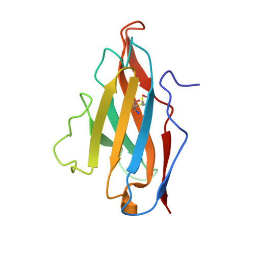
5T93

Immunoglobulin light chain variable domain AL-T05


Experimental Data Snapshot

  • Method: X-RAY DIFFRACTION
  • Resolution: 1.90 Å
  • R-Value Free: 0.223 
  • R-Value Work: 0.171 
  • R-Value Observed: 0.172 

Starting Model: experimental
View more details

wwPDB Validation   3D Report Full Report


This is version 1.6 of the entry. See complete history


Literature

Differences in Protein Concentration Dependence for Nucleation and Elongation in Light Chain Amyloid Formation.

Blancas-Mejia, L.M.Misra, P.Ramirez-Alvarado, M.

(2017) Biochemistry 56: 757-766

  • DOI: https://doi.org/10.1021/acs.biochem.6b01043
  • Primary Citation of Related Structures:  
    5T93

  • PubMed Abstract: 

    Light chain (AL) amyloidosis is a lethal disease characterized by the deposition of the immunoglobulin light chain into amyloid fibrils, resulting in organ dysfunction and failure. Amyloid fibrils have the ability to self-propagate, recruiting soluble protein into the fibril by a nucleation-polymerization mechanism, characteristic of autocatalytic reactions. Experimental data suggest the existence of a critical concentration for initiation of fibril formation. As such, the initial concentration of soluble amyloidogenic protein is expected to have a profound effect on the rate of fibril formation. In this work, we present in vitro evidence that fibril formation rates for AL light chains are affected by the protein concentration in a differential manner. De novo reactions of the proteins with the fastest amyloid kinetics (AL-09, AL-T05, and AL-103) do not present protein concentration dependence. Seeded reactions, however, exhibited weak protein concentration dependence. For AL-12, seeded and protein concentration dependence data suggest a synergistic effect for recruitment and elongation at low protein concentrations, while reactions of κI exhibited poor efficiency in nucleating and elongating preformed fibrils. Additionally, co-aggregation and cross seeding of κI variable domain (V L ) and the κI full length (FL) light chain indicate that the presence of the constant domain in κI FL modulates fibril formation, facilitating the recruitment of κI V L . Together, these results indicate that the dominant process in fibril formation varies among the AL proteins tested with a differential dependence of the protein concentration.


  • Organizational Affiliation

    Department of Biochemistry and Molecular Biology and ‡Department of Immunology, Mayo Clinic , Rochester, Minnesota 55905, United States.


Macromolecules
Find similar proteins by:  (by identity cutoff)  |  3D Structure
Entity ID: 1
MoleculeChains Sequence LengthOrganismDetailsImage
ALT-05 immunoglobulin light chain variable domain from light chain amyloidosis patient
A, B, C, D
110Homo sapiensMutation(s): 0 
Gene Names: IGLV
Entity Groups  
Sequence Clusters30% Identity50% Identity70% Identity90% Identity95% Identity100% Identity
Sequence Annotations
Expand
  • Reference Sequence
Small Molecules
Ligands 2 Unique
IDChains Name / Formula / InChI Key2D Diagram3D Interactions
ZN
Query on ZN

Download Ideal Coordinates CCD File 
E [auth A]
H [auth B]
J [auth B]
M [auth C]
P [auth C]
E [auth A],
H [auth B],
J [auth B],
M [auth C],
P [auth C],
R [auth D],
U [auth D]
ZINC ION
Zn
PTFCDOFLOPIGGS-UHFFFAOYSA-N
ACT
Query on ACT

Download Ideal Coordinates CCD File 
F [auth A]
G [auth A]
I [auth B]
K [auth B]
L [auth B]
F [auth A],
G [auth A],
I [auth B],
K [auth B],
L [auth B],
N [auth C],
O [auth C],
Q [auth C],
S [auth D],
T [auth D]
ACETATE ION
C2 H3 O2
QTBSBXVTEAMEQO-UHFFFAOYSA-M
Experimental Data & Validation

Experimental Data

  • Method: X-RAY DIFFRACTION
  • Resolution: 1.90 Å
  • R-Value Free: 0.223 
  • R-Value Work: 0.171 
  • R-Value Observed: 0.172 
  • Space Group: P 1
Unit Cell:
Length ( Å )Angle ( ˚ )
a = 38.459α = 80.66
b = 42.118β = 86.24
c = 64.995γ = 63.02
Software Package:
Software NamePurpose
PHENIXrefinement
CrystalCleardata reduction
CrystalCleardata scaling
PHASERphasing

Structure Validation

View Full Validation Report


Entry History & Funding Information

Deposition Data


Funding OrganizationLocationGrant Number
National Institutes of Health/National Institute of General Medical Sciences (NIH/NIGMS)United StatesR01 GM 071514

Revision History  (Full details and data files)

  • Version 1.0: 2017-09-13
    Type: Initial release
  • Version 1.1: 2017-09-20
    Changes: Derived calculations
  • Version 1.2: 2018-03-07
    Changes: Data collection
  • Version 1.3: 2018-08-22
    Changes: Data collection, Database references
  • Version 1.4: 2019-12-25
    Changes: Author supporting evidence
  • Version 1.5: 2023-10-04
    Changes: Data collection, Database references, Derived calculations, Refinement description
  • Version 1.6: 2024-11-20
    Changes: Structure summary