5TGC | pdb_00005tgc

Structure of the hetero-trimer of Rtt102-Arp7/9 bound to ATP


Experimental Data Snapshot

  • Method: X-RAY DIFFRACTION
  • Resolution: 3.25 Å
  • R-Value Free: 
    0.322 (Depositor), 0.320 (DCC) 
  • R-Value Work: 
    0.276 (Depositor), 0.280 (DCC) 
  • R-Value Observed: 
    0.278 (Depositor) 

Starting Model: experimental
View more details

wwPDB Validation   3D Report Full Report


Ligand Structure Quality Assessment 

Created with Raphaël 2.3.0Worse 01 BetterLigand structure goodness of fit to experimental dataBest fitted ATPClick on this verticalbar to view details

This is version 1.6 of the entry. See complete history


Literature

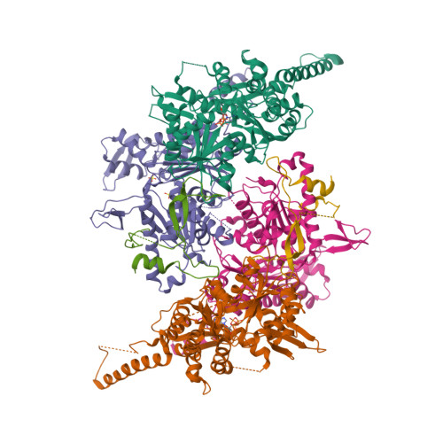
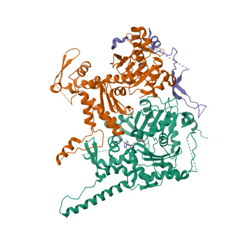
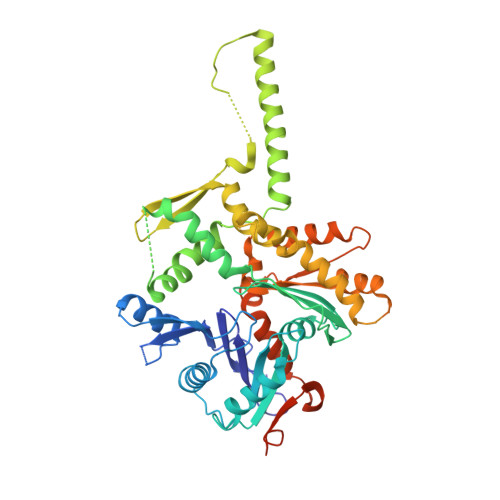
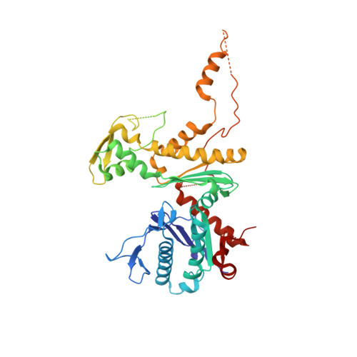
Actin-related proteins regulate the RSC chromatin remodeler by weakening intramolecular interactions of the Sth1 ATPase.

Turegun, B.Baker, R.W.Leschziner, A.E.Dominguez, R.

(2018) Commun Biol 1

  • DOI: https://doi.org/10.1038/s42003-017-0002-6
  • Primary Citation of Related Structures:  
    5TGC

  • PubMed Abstract: 

    The catalytic subunits of SWI/SNF-family and INO80-family chromatin remodelers bind actin and actin-related proteins (Arps) through an N-terminal helicase/SANT-associated (HSA) domain. Between the HSA and ATPase domains lies a conserved post-HSA (pHSA) domain. The HSA domain of Sth1, the catalytic subunit of the yeast SWI/SNF-family remodeler RSC, recruits the Rtt102-Arp7/9 heterotrimer. Rtt102-Arp7/9 regulates RSC function, but the mechanism is unclear. We show that the pHSA domain interacts directly with another conserved region of the catalytic subunit, protrusion-1. Rtt102-Arp7/9 binding to the HSA domain weakens this interaction and promotes the formation of stable, monodisperse complexes with DNA and nucleosomes. A crystal structure of Rtt102-Arp7/9 shows that ATP binds to Arp7 but not Arp9. However, Arp7 does not hydrolyze ATP. Together, the results suggest that Rtt102 and ATP stabilize a conformation of Arp7/9 that potentiates binding to the HSA domain, which releases intramolecular interactions within Sth1 and controls DNA and nucleosome binding.


  • Organizational Affiliation

    Department of Physiology, Perelman School of Medicine, University of Pennsylvania, 728 Clinical Research Building, 415 Curie Boulevard, Philadelphia, PA, 19104-6085, USA.


Macromolecules
Find similar proteins by:  (by identity cutoff)  |  3D Structure
Entity ID: 1
MoleculeChains Sequence LengthOrganismDetailsImage
Actin-related protein 7
A, D
490Saccharomyces cerevisiaeMutation(s): 0 
Gene Names: ARP7
UniProt
Find proteins for Q12406 (Saccharomyces cerevisiae (strain ATCC 204508 / S288c))
Explore Q12406 
Go to UniProtKB:  Q12406
Entity Groups  
Sequence Clusters30% Identity50% Identity70% Identity90% Identity95% Identity100% Identity
UniProt GroupQ12406
Sequence Annotations
Expand
  • Reference Sequence
Find similar proteins by:  (by identity cutoff)  |  3D Structure
Entity ID: 2
MoleculeChains Sequence LengthOrganismDetailsImage
Actin-like protein ARP9
B, E
467Saccharomyces cerevisiaeMutation(s): 0 
Gene Names: ARP9
UniProt
Find proteins for Q05123 (Saccharomyces cerevisiae (strain ATCC 204508 / S288c))
Explore Q05123 
Go to UniProtKB:  Q05123
Entity Groups  
Sequence Clusters30% Identity50% Identity70% Identity90% Identity95% Identity100% Identity
UniProt GroupQ05123
Sequence Annotations
Expand
  • Reference Sequence
Find similar proteins by:  (by identity cutoff)  |  3D Structure
Entity ID: 3
MoleculeChains Sequence LengthOrganismDetailsImage
Regulator of Ty1 transposition protein 102
C, F
158Saccharomyces cerevisiaeMutation(s): 0 
Gene Names: RTT102
UniProt
Find proteins for P53330 (Saccharomyces cerevisiae (strain ATCC 204508 / S288c))
Explore P53330 
Go to UniProtKB:  P53330
Entity Groups  
Sequence Clusters30% Identity50% Identity70% Identity90% Identity95% Identity100% Identity
UniProt GroupP53330
Sequence Annotations
Expand
  • Reference Sequence
Experimental Data & Validation

Experimental Data

  • Method: X-RAY DIFFRACTION
  • Resolution: 3.25 Å
  • R-Value Free:  0.322 (Depositor), 0.320 (DCC) 
  • R-Value Work:  0.276 (Depositor), 0.280 (DCC) 
  • R-Value Observed: 0.278 (Depositor) 
Space Group: P 1
Unit Cell:
Length ( Å )Angle ( ˚ )
a = 79.387α = 109.03
b = 87.982β = 104.64
c = 105.43γ = 96.2
Software Package:
Software NamePurpose
PHENIXrefinement
PDB_EXTRACTdata extraction
SAINTdata scaling

Structure Validation

View Full Validation Report


Ligand Structure Quality Assessment 

Created with Raphaël 2.3.0Worse 01 BetterLigand structure goodness of fit to experimental dataBest fitted ATPClick on this verticalbar to view details

Entry History & Funding Information

Deposition Data


Funding OrganizationLocationGrant Number
National Institutes of Health/National Institute of General Medical Sciences (NIH/NIGMS)United StatesR01_GM073791

Revision History  (Full details and data files)

  • Version 1.0: 2017-09-06
    Type: Initial release
  • Version 1.1: 2017-09-27
    Changes: Author supporting evidence
  • Version 1.2: 2018-03-07
    Changes: Data collection
  • Version 1.3: 2018-03-28
    Changes: Data collection, Database references
  • Version 1.4: 2018-06-13
    Changes: Data collection, Database references
  • Version 1.5: 2019-12-25
    Changes: Author supporting evidence
  • Version 1.6: 2023-10-04
    Changes: Data collection, Database references, Refinement description