5VMC | pdb_00005vmc

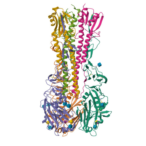
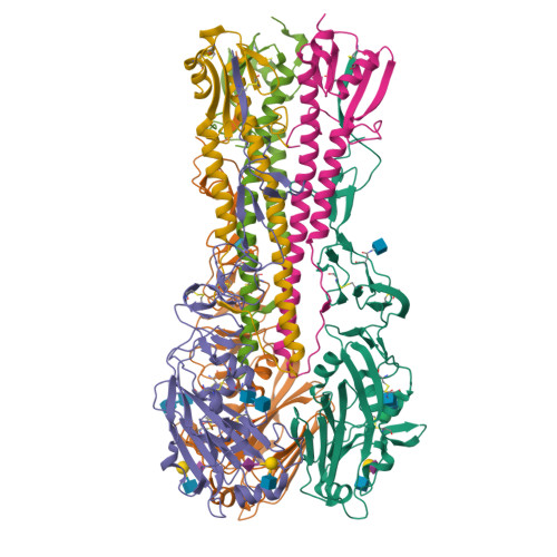
Influenza hemagglutinin H1 mutant DH1 in complex with 6'SLN


Experimental Data Snapshot

  • Method: X-RAY DIFFRACTION
  • Resolution: 2.15 Å
  • R-Value Free: 
    0.205 (Depositor), 0.210 (DCC) 
  • R-Value Work: 
    0.176 (Depositor), 0.180 (DCC) 
  • R-Value Observed: 
    0.177 (Depositor) 

Starting Model: experimental
View more details

wwPDB Validation   3D Report Full Report


Ligand Structure Quality Assessment 

Created with Raphaël 2.3.0Worse 01 BetterLigand structure goodness of fit to experimental dataBest fitted NAGClick on this verticalbar to view details

This is version 2.2 of the entry. See complete history


Literature

Determinant of receptor-preference switch in influenza hemagglutinin.

Ni, F.Kondrashkina, E.Wang, Q.

(2017) Virology 513: 98-107

  • DOI: https://doi.org/10.1016/j.virol.2017.10.010
  • Primary Citation of Related Structures:  
    5VMC, 5VMF, 5VMG, 5VMJ

  • PubMed Abstract: 

    Influenza pandemic occurs when a new strain from other animal species overcomes the inter-species barriers and supports rapid human-to-human transmission. A critical prerequisite to this process is that hemagglutinin (HA) acquires a few key mutations to switch from avian receptors to human receptors. Previous studies suggest that H1 and H2/H3 HAs use different sets of mutations for the switch. This report shows that HA from the 1918 H1N1 pandemic virus (1918H1 HA) adopts the set of mutations used by H2/H3 HAs in receptor-preference switch when its 130-loop is made similar to those of H2/H3 HAs. Thus, the 130-loop appears to be the key determinant for the different mutations employed by pandemic H1 or H2/H3 HA. The correlation of the mutational routes and the 130-loop as unraveled in this study opens the door for efficient investigation of mutations required by other HA subtypes for inter-human airborne transmission.


  • Organizational Affiliation

    Verna and Marrs McLean Department of Biochemistry and Molecular Biology, Baylor College of Medicine, One Baylor Plaza, Houston, TX 77030, USA.


Macromolecules
Find similar proteins by:  (by identity cutoff)  |  3D Structure
Entity ID: 1
MoleculeChains Sequence LengthOrganismDetailsImage
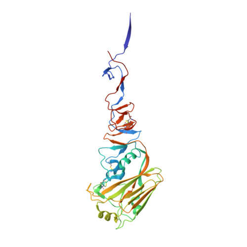
Hemagglutinin HA1
A, C, E
326Influenza A virus (A/Brevig Mission/1/1918(H1N1))Mutation(s): 0 
Gene Names: HA
UniProt
Find proteins for Q9WFX3 (Influenza A virus (strain A/Brevig Mission/1/1918 H1N1))
Explore Q9WFX3 
Go to UniProtKB:  Q9WFX3
Entity Groups  
Sequence Clusters30% Identity50% Identity70% Identity90% Identity95% Identity100% Identity
UniProt GroupQ9WFX3
Glycosylation
Glycosylation Sites: 2
Sequence Annotations
Expand
  • Reference Sequence
Find similar proteins by:  (by identity cutoff)  |  3D Structure
Entity ID: 2
MoleculeChains Sequence LengthOrganismDetailsImage
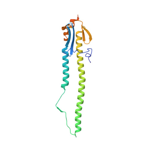
Hemagglutinin HA2
B, D, F
191Influenza A virus (A/Brevig Mission/1/1918(H1N1))Mutation(s): 0 
Gene Names: HA
UniProt
Find proteins for Q9WFX3 (Influenza A virus (strain A/Brevig Mission/1/1918 H1N1))
Explore Q9WFX3 
Go to UniProtKB:  Q9WFX3
Entity Groups  
Sequence Clusters30% Identity50% Identity70% Identity90% Identity95% Identity100% Identity
UniProt GroupQ9WFX3
Sequence Annotations
Expand
  • Reference Sequence
Oligosaccharides

Help

Entity ID: 3
MoleculeChains Length2D Diagram Glycosylation3D Interactions
beta-D-mannopyranose-(1-4)-2-acetamido-2-deoxy-beta-D-glucopyranose-(1-4)-2-acetamido-2-deoxy-beta-D-glucopyranose
G
3N-Glycosylation
Glycosylation Resources
GlyTouCan:  G15407YE
GlyCosmos:  G15407YE
GlyGen:  G15407YE
Entity ID: 4
MoleculeChains Length2D Diagram Glycosylation3D Interactions
N-acetyl-alpha-neuraminic acid-(2-6)-beta-D-galactopyranose-(1-4)-2-acetamido-2-deoxy-beta-D-glucopyranose
H, J, L
3N/A
Glycosylation Resources
GlyTouCan:  G73578JC
GlyCosmos:  G73578JC
GlyGen:  G73578JC
Entity ID: 5
MoleculeChains Length2D Diagram Glycosylation3D Interactions
2-acetamido-2-deoxy-beta-D-glucopyranose-(1-4)-2-acetamido-2-deoxy-beta-D-glucopyranose
I, K
2N-Glycosylation
Glycosylation Resources
GlyTouCan:  G42666HT
GlyCosmos:  G42666HT
GlyGen:  G42666HT
Experimental Data & Validation

Experimental Data

  • Method: X-RAY DIFFRACTION
  • Resolution: 2.15 Å
  • R-Value Free:  0.205 (Depositor), 0.210 (DCC) 
  • R-Value Work:  0.176 (Depositor), 0.180 (DCC) 
  • R-Value Observed: 0.177 (Depositor) 
Space Group: P 1 21 1
Unit Cell:
Length ( Å )Angle ( ˚ )
a = 95.68α = 90
b = 81.82β = 90.79
c = 121.46γ = 90
Software Package:
Software NamePurpose
PHENIXrefinement
MOSFLMdata reduction
SCALAdata scaling
PHENIXphasing

Structure Validation

View Full Validation Report


Ligand Structure Quality Assessment 

Created with Raphaël 2.3.0Worse 01 BetterLigand structure goodness of fit to experimental dataBest fitted NAGClick on this verticalbar to view details

Entry History 

Deposition Data

Revision History  (Full details and data files)

  • Version 1.0: 2017-11-08
    Type: Initial release
  • Version 2.0: 2020-07-29
    Type: Remediation
    Reason: Carbohydrate remediation
    Changes: Advisory, Atomic model, Data collection, Derived calculations, Structure summary
  • Version 2.1: 2023-10-04
    Changes: Data collection, Database references, Derived calculations, Refinement description, Structure summary
  • Version 2.2: 2024-10-09
    Changes: Structure summary