5Y7R

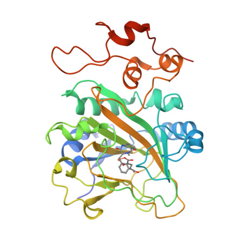
Quaternary complex of AsqJ-Fe3+-2OG-cyclopeptin


Experimental Data Snapshot

  • Method: X-RAY DIFFRACTION
  • Resolution: 1.96 Å
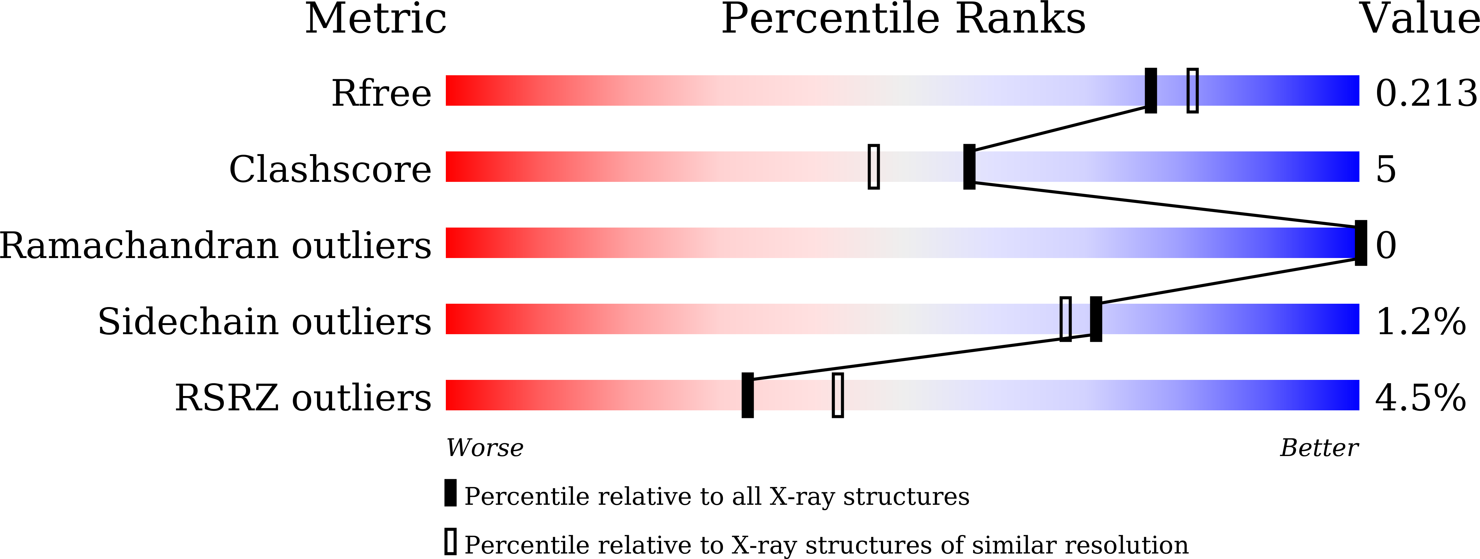
  • R-Value Free: 0.215 
  • R-Value Work: 0.166 
  • R-Value Observed: 0.168 

wwPDB Validation   3D Report Full Report


Ligand Structure Quality Assessment 


This is version 1.3 of the entry. See complete history


Literature

Insights into the Desaturation of Cyclopeptin and its C3 Epimer Catalyzed by a non-Heme Iron Enzyme: Structural Characterization and Mechanism Elucidation.

Liao, H.J.Li, J.Huang, J.L.Davidson, M.Kurnikov, I.Lin, T.S.Lee, J.L.Kurnikova, M.Guo, Y.Chan, N.L.Chang, W.C.

(2018) Angew Chem Int Ed Engl 57: 1831-1835

  • DOI: https://doi.org/10.1002/anie.201710567
  • Primary Citation of Related Structures:  
    5Y7R, 5Y7T

  • PubMed Abstract: 

    AsqJ, an iron(II)- and 2-oxoglutarate-dependent enzyme found in viridicatin-type alkaloid biosynthetic pathways, catalyzes sequential desaturation and epoxidation to produce cyclopenins. Crystal structures of AsqJ bound to cyclopeptin and its C3 epimer are reported. Meanwhile, a detailed mechanistic study was carried out to decipher the desaturation mechanism. These findings suggest that a pathway involving hydrogen atom abstraction at the C10 position of the substrate by a short-lived Fe IV -oxo species and the subsequent formation of a carbocation or a hydroxylated intermediate is preferred during AsqJ-catalyzed desaturation.


  • Organizational Affiliation

    Institute of Biochemistry and Molecular Biology, College of Medicine, National (Taiwan) University, Taipei, 100, Taiwan.


Macromolecules
Find similar proteins by:  (by identity cutoff)  |  3D Structure
Entity ID: 1
MoleculeChains Sequence LengthOrganismDetailsImage
Iron/alpha-ketoglutarate-dependent dioxygenase asqJA [auth B]308Aspergillus nidulans FGSC A4Mutation(s): 0 
Gene Names: asqJAN9227
EC: 1.14
UniProt
Find proteins for Q5AR53 (Emericella nidulans (strain FGSC A4 / ATCC 38163 / CBS 112.46 / NRRL 194 / M139))
Go to UniProtKB:  Q5AR53
Sequence Annotations
Expand
  • Reference Sequence
Experimental Data & Validation

Experimental Data

  • Method: X-RAY DIFFRACTION
  • Resolution: 1.96 Å
  • R-Value Free: 0.215 
  • R-Value Work: 0.166 
  • R-Value Observed: 0.168 
  • Space Group: C 2 2 21
Unit Cell:
Length ( Å )Angle ( ˚ )
a = 73.441α = 90
b = 119.689β = 90
c = 66.207γ = 90
Software Package:
Software NamePurpose
PHENIXrefinement
HKL-2000data scaling

Structure Validation

View Full Validation Report


Ligand Structure Quality Assessment 


Entry History 

Deposition Data

Revision History  (Full details and data files)

  • Version 1.0: 2018-01-24
    Type: Initial release
  • Version 1.1: 2018-01-31
    Changes: Database references
  • Version 1.2: 2018-02-21
    Changes: Database references
  • Version 1.3: 2024-03-27
    Changes: Data collection, Database references, Derived calculations