6DP6

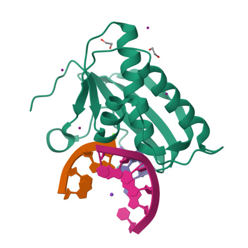
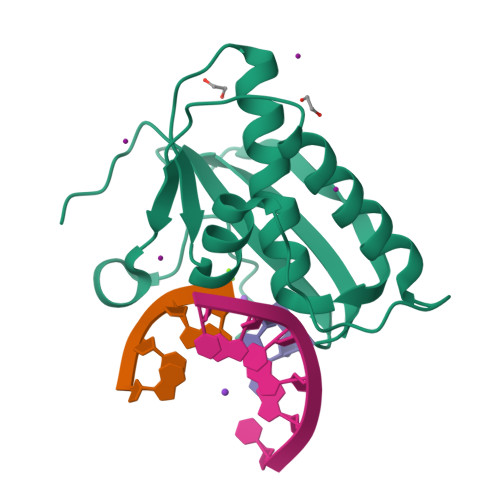
Crystal Structure of Bacillus Halodurans Ribonuclease H1 in Complex with an RNA/DNA Hybrid: Reaction in 80 mM Mg2+ and 75 mM K+ for 40 s at 21 C


Experimental Data Snapshot

  • Method: X-RAY DIFFRACTION
  • Resolution: 1.40 Å
  • R-Value Free: 0.186 
  • R-Value Work: 0.163 
  • R-Value Observed: 0.164 

Starting Model: experimental
View more details

wwPDB Validation   3D Report Full Report


This is version 1.2 of the entry. See complete history


Literature

Cation trafficking propels RNA hydrolysis.

Samara, N.L.Yang, W.

(2018) Nat Struct Mol Biol 25: 715-721

  • DOI: https://doi.org/10.1038/s41594-018-0099-4
  • Primary Citation of Related Structures:  
    6DMN, 6DMV, 6DO8, 6DO9, 6DOA, 6DOB, 6DOC, 6DOD, 6DOE, 6DOF, 6DOG, 6DOH, 6DOI, 6DOJ, 6DOK, 6DOL, 6DOM, 6DON, 6DOO, 6DOP, 6DOQ, 6DOR, 6DOS, 6DOT, 6DOU, 6DOV, 6DOW, 6DOX, 6DOY, 6DOZ, 6DP0, 6DP1, 6DP2, 6DP3, 6DP4, 6DP5, 6DP6, 6DP7, 6DP8, 6DP9, 6DPA, 6DPB, 6DPC, 6DPD, 6DPE, 6DPF, 6DPG, 6DPH, 6DPI, 6DPJ

  • PubMed Abstract: 

    Catalysis by members of the RNase H superfamily of enzymes is generally believed to require only two Mg 2+ ions that are coordinated by active-site carboxylates. By examining the catalytic process of Bacillus halodurans RNase H1 in crystallo, however, we found that the two canonical Mg 2+ ions and an additional K + failed to align the nucleophilic water for RNA cleavage. Substrate alignment and product formation required a second K + and a third Mg 2+ , which replaced the first K + and departed immediately after cleavage. A third transient Mg 2+ has also been observed for DNA synthesis, but in that case it coordinates the leaving group instead of the nucleophile as in the case of the RNase H1 hydrolysis reaction. These transient cations have no contact with the enzymes. Other DNA and RNA enzymes that catalyze consecutive cleavage and strand-transfer reactions in a single active site may similarly require cation trafficking coordinated by the substrate.


  • Organizational Affiliation

    Laboratory of Molecular Biology, National Institute of Diabetes and Digestive and Kidney Diseases (NIDDK), US National Institutes of Health (NIH), Bethesda, MD, USA.


Macromolecules

Find similar proteins by:  (by identity cutoff)  |  3D Structure
Entity ID: 1
MoleculeChains Sequence LengthOrganismDetailsImage
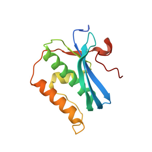
Ribonuclease H142Halalkalibacterium halodurans C-125Mutation(s): 0 
Gene Names: rnhABH0863
EC: 3.1.26.4
UniProt
Find proteins for Q9KEI9 (Halalkalibacterium halodurans (strain ATCC BAA-125 / DSM 18197 / FERM 7344 / JCM 9153 / C-125))
Explore Q9KEI9 
Go to UniProtKB:  Q9KEI9
Entity Groups  
Sequence Clusters30% Identity50% Identity70% Identity90% Identity95% Identity100% Identity
UniProt GroupQ9KEI9
Sequence Annotations
Expand
  • Reference Sequence

Find similar nucleic acids by:  Sequence   |   3D Structure  

Entity ID: 2
MoleculeChains LengthOrganismImage
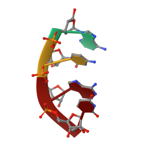
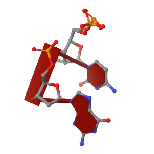
5'-R(*AP*CP*AP*U)-3' portion of cleaved RNA 5'-R(*AP*CP*AP*UP*CP*G)-3'4synthetic construct
Sequence Annotations
Expand
  • Reference Sequence

Find similar nucleic acids by:  Sequence   |   3D Structure  

Entity ID: 3
MoleculeChains LengthOrganismImage
5'-R(P*CP*G)-3' portion of cleaved RNA 5'-R(*AP*CP*AP*UP*CP*G)-3'C [auth b]2synthetic construct
Sequence Annotations
Expand
  • Reference Sequence

Find similar nucleic acids by:  Sequence   |   3D Structure  

Entity ID: 4
MoleculeChains LengthOrganismImage
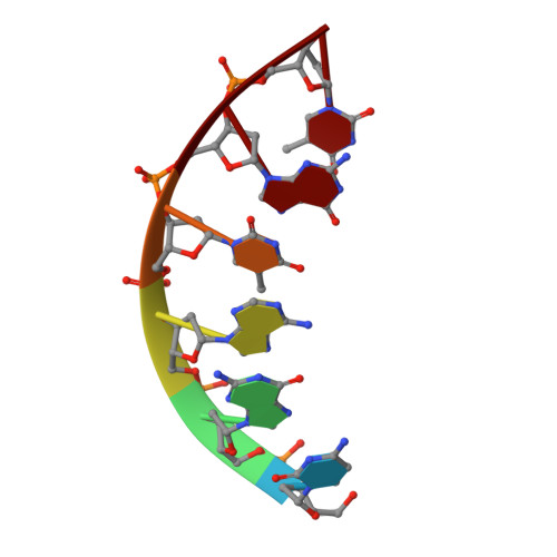
DNA (5'-D(*CP*GP*AP*TP*GP*T)-3')D [auth C]6synthetic construct
Sequence Annotations
Expand
  • Reference Sequence
Small Molecules
Ligands 4 Unique
IDChains Name / Formula / InChI Key2D Diagram3D Interactions
IOD
Query on IOD

Download Ideal Coordinates CCD File 
I [auth A],
J [auth A],
K [auth A],
L [auth A]
IODIDE ION
I
XMBWDFGMSWQBCA-UHFFFAOYSA-M
EDO
Query on EDO

Download Ideal Coordinates CCD File 
M [auth A],
N [auth A]
1,2-ETHANEDIOL
C2 H6 O2
LYCAIKOWRPUZTN-UHFFFAOYSA-N
K (Subject of Investigation/LOI)
Query on K

Download Ideal Coordinates CCD File 
G [auth A],
H [auth A],
O [auth C]
POTASSIUM ION
K
NPYPAHLBTDXSSS-UHFFFAOYSA-N
MG (Subject of Investigation/LOI)
Query on MG

Download Ideal Coordinates CCD File 
E [auth A],
F [auth A]
MAGNESIUM ION
Mg
JLVVSXFLKOJNIY-UHFFFAOYSA-N
Experimental Data & Validation

Experimental Data

  • Method: X-RAY DIFFRACTION
  • Resolution: 1.40 Å
  • R-Value Free: 0.186 
  • R-Value Work: 0.163 
  • R-Value Observed: 0.164 
  • Space Group: C 1 2 1
Unit Cell:
Length ( Å )Angle ( ˚ )
a = 81.832α = 90
b = 37.292β = 96.71
c = 62.607γ = 90
Software Package:
Software NamePurpose
Aimlessdata scaling
PHENIXrefinement
PDB_EXTRACTdata extraction
HKL-2000data reduction
PHENIXphasing

Structure Validation

View Full Validation Report


Entry History 

Deposition Data

Revision History  (Full details and data files)

  • Version 1.0: 2018-08-15
    Type: Initial release
  • Version 1.1: 2018-08-22
    Changes: Data collection, Database references
  • Version 1.2: 2023-10-11
    Changes: Data collection, Database references, Derived calculations, Refinement description