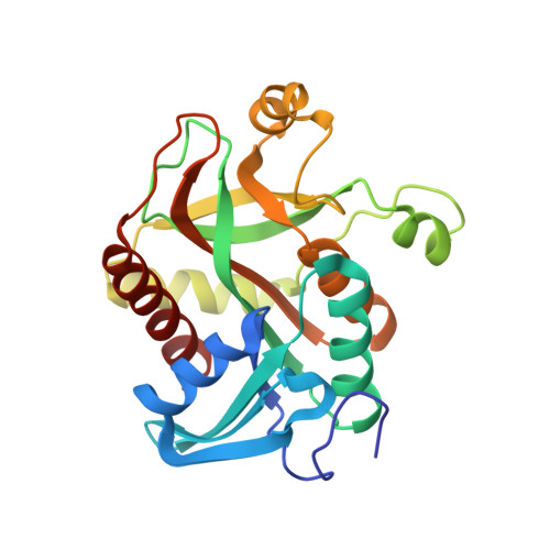
Helicobacter pylori purine nucleoside phosphorylase shows new distribution patterns of open and closed active site conformations and unusual biochemical features.
Narczyk, M., Bertosa, B., Papa, L., Vukovic, V., Lescic Asler, I., Wielgus-Kutrowska, B., Bzowska, A., Luic, M., Stefanic, Z.(2018) FEBS J 285: 1305-1325
- PubMed: 29430816
- DOI: https://doi.org/10.1111/febs.14403
- Primary Citation of Related Structures:
5LU0, 6F4W, 6F4X, 6F52, 6F5A, 6F5I - PubMed Abstract:
Even with decades of research, purine nucleoside phosphorylases (PNPs) are enzymes whose mechanism is yet to be fully understood. This is especially true in the case of hexameric PNPs, and is probably, in part, due to their complex oligomeric nature and a whole spectrum of active site conformations related to interactions with different ligands. Here we report an extensive structural characterization of the apo forms of hexameric PNP from Helicobacter pylori (HpPNP), as well as its complexes with phosphate (P i ) and an inhibitor, formycin A (FA), together with kinetic, binding, docking and molecular dynamics studies. X-ray structures show previously unseen distributions of open and closed active sites. Microscale thermophoresis results indicate that a two-site model describes P i binding, while a three-site model is needed to characterize FA binding, irrespective of P i presence. The latter may be related to the newly found nonstandard mode of FA binding. The ternary complex of the enzyme with P i and FA shows, however, that P i binding stabilizes the standard mode of FA binding. Surprisingly, HpPNP has low affinity towards the natural substrate adenosine. Molecular dynamics simulations show that P i moves out of most active sites, in accordance with its weak binding. Conformational changes between nonstandard and standard binding modes of nucleoside are observed during the simulations. Altogether, these findings show some unique features of HpPNP and provide new insights into the functioning of the active sites, with implications for understanding the complex mechanism of catalysis of this enzyme. The atomic coordinates and structure factors have been deposited in the Protein Data Bank: with accession codes 6F52 (HpPNPapo_1), 6F5A (HpPNPapo_2), 6F5I (HpPNPapo_3), 5LU0 (HpPNP_PO4), 6F4W (HpPNP_FA) and 6F4X (HpPNP_PO4_FA). Purine nucleoside orthophosphate ribosyl transferase, EC2.4.2.1, UniProtID: P56463.
Organizational Affiliation:
Division of Biophysics, Institute of Experimental Physics, Faculty of Physics, University of Warsaw, Poland.


