6GM5

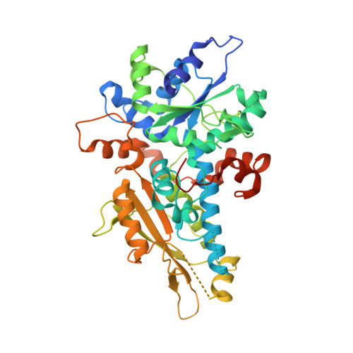
[FeFe]-hydrogenase HydA1 from Chlamydomonas reinhardtii,variant E141A


Experimental Data Snapshot

  • Method: X-RAY DIFFRACTION
  • Resolution: 1.45 Å
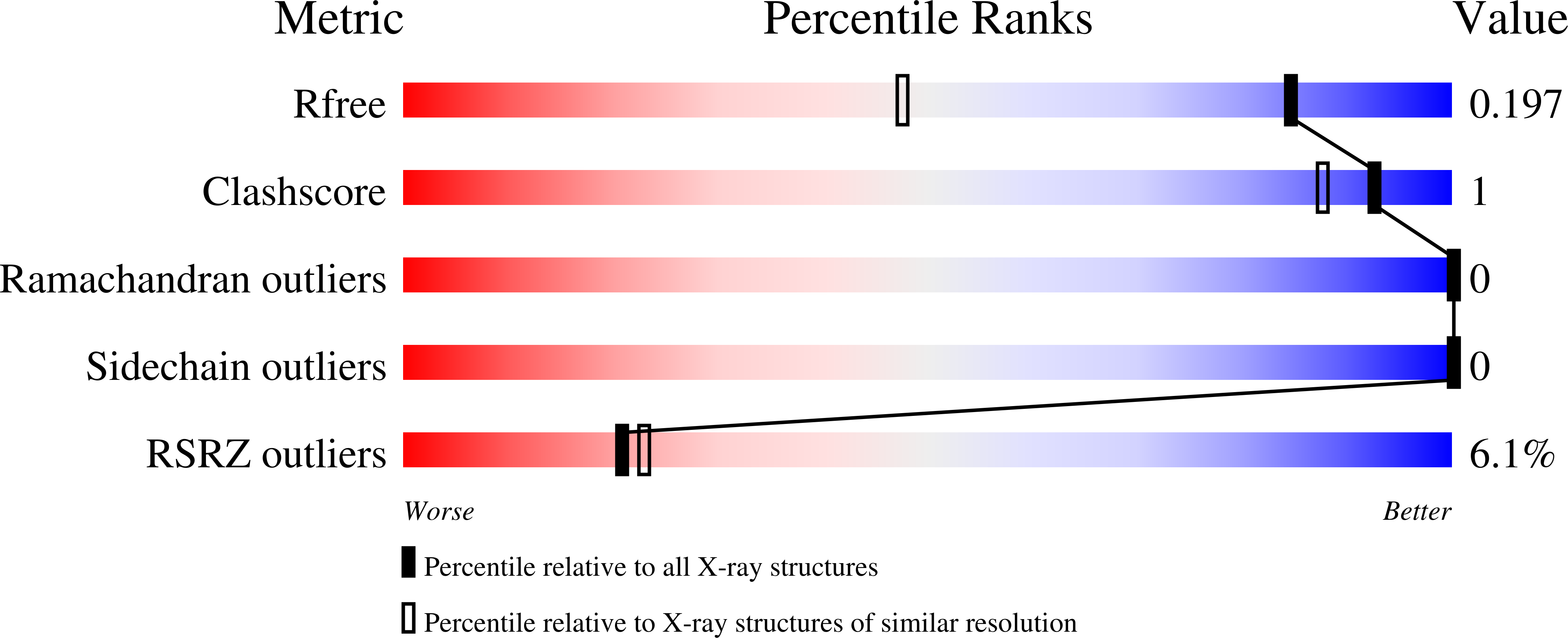
  • R-Value Free: 0.197 
  • R-Value Work: 0.165 
  • R-Value Observed: 0.167 

Starting Model: experimental
View more details

wwPDB Validation   3D Report Full Report


This is version 1.3 of the entry. See complete history


Literature

Crystallographic and spectroscopic assignment of the proton transfer pathway in [FeFe]-hydrogenases.

Duan, J.Senger, M.Esselborn, J.Engelbrecht, V.Wittkamp, F.Apfel, U.P.Hofmann, E.Stripp, S.T.Happe, T.Winkler, M.

(2018) Nat Commun 9: 4726-4726

  • DOI: https://doi.org/10.1038/s41467-018-07140-x
  • Primary Citation of Related Structures:  
    6GLY, 6GLZ, 6GM0, 6GM1, 6GM2, 6GM3, 6GM4, 6GM5, 6GM6, 6GM7, 6GM8

  • PubMed Abstract: 

    The unmatched catalytic turnover rates of [FeFe]-hydrogenases require an exceptionally efficient proton-transfer (PT) pathway to shuttle protons as substrates or products between bulk water and catalytic center. For clostridial [FeFe]-hydrogenase CpI such a pathway has been proposed and analyzed, but mainly on a theoretical basis. Here, eleven enzyme variants of two different [FeFe]-hydrogenases (CpI and HydA1) with substitutions in the presumptive PT-pathway are examined kinetically, spectroscopically, and crystallographically to provide solid experimental proof for its role in hydrogen-turnover. Targeting key residues of the PT-pathway by site directed mutagenesis significantly alters the pH-activity profile of these variants and in presence of H 2 their cofactor is trapped in an intermediate state indicative of precluded proton-transfer. Furthermore, crystal structures coherently explain the individual levels of residual activity, demonstrating e.g. how trapped H 2 O molecules rescue the interrupted PT-pathway. These features provide conclusive evidence that the targeted positions are indeed vital for catalytic proton-transfer.


  • Organizational Affiliation

    Department of Plant Biochemistry, Photobiotechnology, Ruhr-Universität Bochum, 44801, Bochum, Germany.


Macromolecules
Find similar proteins by:  (by identity cutoff)  |  3D Structure
Entity ID: 1
MoleculeChains Sequence LengthOrganismDetailsImage
Fe-hydrogenase452Chlamydomonas reinhardtiiMutation(s): 1 
Gene Names: hyd1HYD1hydAhydA1CHLRE_03g199800v5CHLREDRAFT_183963
EC: 1.18.99.1
UniProt
Find proteins for Q9FYU1 (Chlamydomonas reinhardtii)
Explore Q9FYU1 
Go to UniProtKB:  Q9FYU1
Entity Groups  
Sequence Clusters30% Identity50% Identity70% Identity90% Identity95% Identity100% Identity
UniProt GroupQ9FYU1
Sequence Annotations
Expand
  • Reference Sequence
Experimental Data & Validation

Experimental Data

  • Method: X-RAY DIFFRACTION
  • Resolution: 1.45 Å
  • R-Value Free: 0.197 
  • R-Value Work: 0.165 
  • R-Value Observed: 0.167 
  • Space Group: P 32 2 1
Unit Cell:
Length ( Å )Angle ( ˚ )
a = 70.62α = 90
b = 70.62β = 90
c = 154.91γ = 120
Software Package:
Software NamePurpose
PHENIXrefinement
XSCALEdata scaling
PDB_EXTRACTdata extraction
XDSdata reduction
PHENIXphasing

Structure Validation

View Full Validation Report


Entry History 

Deposition Data

Revision History  (Full details and data files)

  • Version 1.0: 2018-11-07
    Type: Initial release
  • Version 1.1: 2018-11-21
    Changes: Data collection, Database references
  • Version 1.2: 2019-11-06
    Changes: Data collection, Database references
  • Version 1.3: 2024-01-17
    Changes: Advisory, Data collection, Database references, Derived calculations, Refinement description