6H44

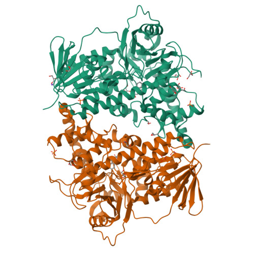
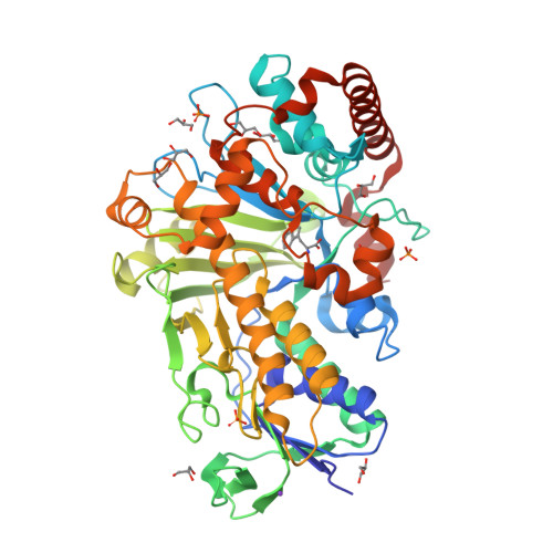
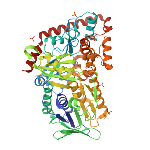
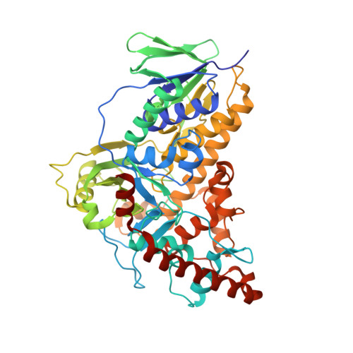
Flavin-dependent Tryptophan 6-halogenase Thal in complex with tryptophan


Experimental Data Snapshot

  • Method: X-RAY DIFFRACTION
  • Resolution: 2.55 Å
  • R-Value Free: 0.225 
  • R-Value Work: 0.179 
  • R-Value Observed: 0.181 

Starting Model: other
View more details

wwPDB Validation   3D Report Full Report


Ligand Structure Quality Assessment 


This is version 1.2 of the entry. See complete history


Literature

Structure-based switch of regioselectivity in the flavin-dependent tryptophan 6-halogenase Thal.

Moritzer, A.C.Minges, H.Prior, T.Frese, M.Sewald, N.Niemann, H.H.

(2019) J Biol Chem 294: 2529-2542

  • DOI: https://doi.org/10.1074/jbc.RA118.005393
  • Primary Citation of Related Structures:  
    6H43, 6H44, 6IB5

  • PubMed Abstract: 

    Flavin-dependent halogenases increasingly attract attention as biocatalysts in organic synthesis, facilitating environmentally friendly halogenation strategies that require only FADH 2 , oxygen, and halide salts. Different flavin-dependent tryptophan halogenases regioselectively chlorinate or brominate trypto-phan's indole moiety at C5, C6, or C7. Here, we present the first substrate-bound structure of a tryptophan 6-halogenase, namely Thal, also known as ThdH, from the bacterium Streptomyces albogriseolus at 2.55 Å resolution. The structure revealed that the C6 of tryptophan is positioned next to the ϵ-amino group of a conserved lysine, confirming the hypothesis that proximity to the catalytic residue determines the site of electrophilic aromatic substitution. Although Thal is more similar in sequence and structure to the tryptophan 7-halogenase RebH than to the tryptophan 5-halogenase PyrH, the indole binding pose in the Thal active site more closely resembled that of PyrH than that of RebH. The difference in indole orientation between Thal and RebH appeared to be largely governed by residues positioning the Trp backbone atoms. The sequences of Thal and RebH lining the substrate binding site differ in only few residues. Therefore, we exchanged five amino acids in the Thal active site with the corresponding counterparts in RebH, generating the quintuple variant Thal-RebH5. Overall conversion of l-Trp by the Thal-RebH5 variant resembled that of WT Thal, but its regioselectivity of chlorination and bromination was almost completely switched from C6 to C7 as in RebH. We conclude that structure-based protein engineering with targeted substitution of a few residues is an efficient approach to tailoring flavin-dependent halogenases.


  • Organizational Affiliation

    From the Structural Biochemistry and.


Macromolecules
Find similar proteins by:  (by identity cutoff)  |  3D Structure
Entity ID: 1
MoleculeChains Sequence LengthOrganismDetailsImage
Tryptophan 6-halogenase
A, B
534Streptomyces albogriseolusMutation(s): 0 
Gene Names: thalthdH
EC: 1.14.19.59
UniProt
Find proteins for A1E280 (Streptomyces albogriseolus)
Explore A1E280 
Go to UniProtKB:  A1E280
Entity Groups  
Sequence Clusters30% Identity50% Identity70% Identity90% Identity95% Identity100% Identity
UniProt GroupA1E280
Sequence Annotations
Expand
  • Reference Sequence
Small Molecules
Ligands 4 Unique
IDChains Name / Formula / InChI Key2D Diagram3D Interactions
TRP
Query on TRP

Download Ideal Coordinates CCD File 
O [auth A],
Z [auth B]
TRYPTOPHAN
C11 H12 N2 O2
QIVBCDIJIAJPQS-VIFPVBQESA-N
PO4
Query on PO4

Download Ideal Coordinates CCD File 
C [auth A]
D [auth A]
E [auth A]
Q [auth B]
R [auth B]
C [auth A],
D [auth A],
E [auth A],
Q [auth B],
R [auth B],
S [auth B],
T [auth B]
PHOSPHATE ION
O4 P
NBIIXXVUZAFLBC-UHFFFAOYSA-K
GOL
Query on GOL

Download Ideal Coordinates CCD File 
F [auth A]
G [auth A]
H [auth A]
I [auth A]
J [auth A]
F [auth A],
G [auth A],
H [auth A],
I [auth A],
J [auth A],
K [auth A],
L [auth A],
M [auth A],
N [auth A],
U [auth B],
V [auth B],
W [auth B],
X [auth B],
Y [auth B]
GLYCEROL
C3 H8 O3
PEDCQBHIVMGVHV-UHFFFAOYSA-N
K
Query on K

Download Ideal Coordinates CCD File 
P [auth A]POTASSIUM ION
K
NPYPAHLBTDXSSS-UHFFFAOYSA-N
Experimental Data & Validation

Experimental Data

  • Method: X-RAY DIFFRACTION
  • Resolution: 2.55 Å
  • R-Value Free: 0.225 
  • R-Value Work: 0.179 
  • R-Value Observed: 0.181 
  • Space Group: P 64
Unit Cell:
Length ( Å )Angle ( ˚ )
a = 136.7α = 90
b = 136.7β = 90
c = 144.9γ = 120
Software Package:
Software NamePurpose
REFMACrefinement
XDSdata reduction
XSCALEdata scaling
PHASERphasing

Structure Validation

View Full Validation Report


Ligand Structure Quality Assessment 


Entry History 

Deposition Data

Revision History  (Full details and data files)

  • Version 1.0: 2018-12-26
    Type: Initial release
  • Version 1.1: 2019-02-27
    Changes: Data collection, Database references
  • Version 1.2: 2024-05-01
    Changes: Data collection, Database references, Refinement description