Structure-based design of selective, histone substrate-based inhibitors of histone lysine demethylases 4 (KDM4) subfamily
Maw, J., Le Bihan, Y.V., Bavetsias, V., Blagg, J.To be published.
Experimental Data Snapshot
Starting Model: experimental
View more details
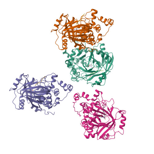
Entity ID: 1 | |||||
|---|---|---|---|---|---|
| Molecule | Chains | Sequence Length | Organism | Details | Image |
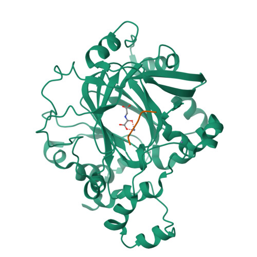
| Lysine-specific demethylase 4A | 360 | Homo sapiens | Mutation(s): 0 Gene Names: KDM4A, JHDM3A, JMJD2, JMJD2A, KIAA0677 EC: 1.14.11 (PDB Primary Data), 1.14.11.66 (UniProt), 1.14.11.69 (UniProt) | 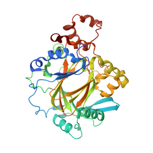 | |
UniProt & NIH Common Fund Data Resources | |||||
Find proteins for O75164 (Homo sapiens) Explore O75164 Go to UniProtKB: O75164 | |||||
PHAROS: O75164 GTEx: ENSG00000066135 | |||||
Entity Groups | |||||
| Sequence Clusters | 30% Identity50% Identity70% Identity90% Identity95% Identity100% Identity | ||||
| UniProt Group | O75164 | ||||
Sequence AnnotationsExpand | |||||
| |||||
Find similar proteins by: Sequence | 3D Structure
Entity ID: 2 | |||||
|---|---|---|---|---|---|
| Molecule | Chains | Sequence Length | Organism | Details | Image |
| Histone H3.3 | 15 | Homo sapiens | Mutation(s): 1 Gene Names: H3F3A, H3.3A, H3F3, PP781, H3F3B, H3.3B | 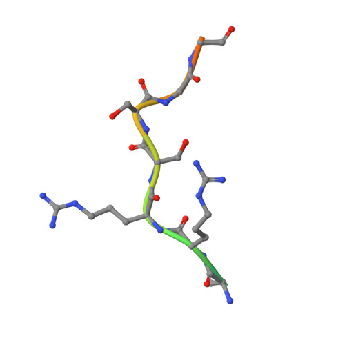 | |
UniProt & NIH Common Fund Data Resources | |||||
Find proteins for P68431 (Homo sapiens) Explore P68431 Go to UniProtKB: P68431 | |||||
PHAROS: P68431 | |||||
Entity Groups | |||||
| Sequence Clusters | 30% Identity50% Identity70% Identity90% Identity95% Identity100% Identity | ||||
| UniProt Group | P68431 | ||||
Sequence AnnotationsExpand | |||||
| |||||
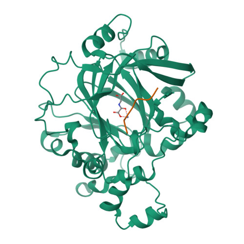
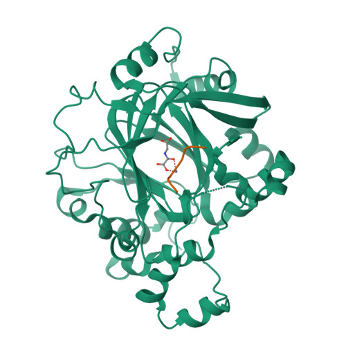
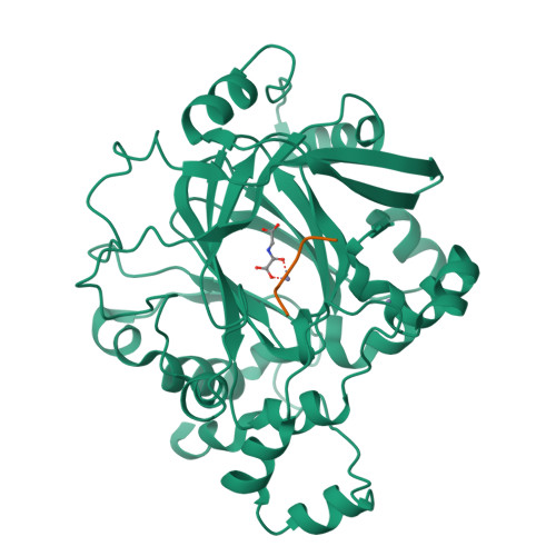
| Ligands 2 Unique | |||||
|---|---|---|---|---|---|
| ID | Chains | Name / Formula / InChI Key | 2D Diagram | 3D Interactions | |
| OGA (Subject of Investigation/LOI) Query on OGA | K [auth A], N [auth B], Q [auth C], T [auth D] | N-OXALYLGLYCINE C4 H5 N O5 BIMZLRFONYSTPT-UHFFFAOYSA-N |  | ||
| ZN Query on ZN | I [auth A] J [auth A] L [auth B] M [auth B] O [auth C] | ZINC ION Zn PTFCDOFLOPIGGS-UHFFFAOYSA-N |  | ||
| Length ( Å ) | Angle ( ˚ ) |
|---|---|
| a = 58.44 | α = 90 |
| b = 103.41 | β = 99.63 |
| c = 144.52 | γ = 90 |
| Software Name | Purpose |
|---|---|
| BUSTER | refinement |
| XDS | data reduction |
| Aimless | data scaling |
| PHASER | phasing |
| Funding Organization | Location | Grant Number |
|---|---|---|
| Cancer Research UK | United Kingdom | C347/A18364 |