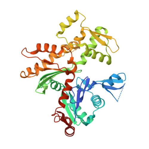
6I4L

Crystal Structure of Plasmodium falciparum actin I (G115A mutant) in the Mg-K-ATP/ADP state


Experimental Data Snapshot

  • Method: X-RAY DIFFRACTION
  • Resolution: 1.83 Å
  • R-Value Free: 0.243 
  • R-Value Work: 0.200 
  • R-Value Observed: 0.202 

Starting Model: experimental
View more details

wwPDB Validation   3D Report Full Report


Ligand Structure Quality Assessment 


This is version 1.1 of the entry. See complete history


Literature

Atomic view into Plasmodium actin polymerization, ATP hydrolysis, and fragmentation.

Kumpula, E.P.Lopez, A.J.Tajedin, L.Han, H.Kursula, I.

(2019) PLoS Biol 17: e3000315-e3000315

  • DOI: https://doi.org/10.1371/journal.pbio.3000315
  • Primary Citation of Related Structures:  
    6I4D, 6I4E, 6I4F, 6I4G, 6I4H, 6I4I, 6I4J, 6I4K, 6I4L, 6I4M

  • PubMed Abstract: 

    Plasmodium actins form very short filaments and have a noncanonical link between ATP hydrolysis and polymerization. Long filaments are detrimental to the parasites, but the structural factors constraining Plasmodium microfilament lengths have remained unknown. Using high-resolution crystallography, we show that magnesium binding causes a slight flattening of the Plasmodium actin I monomer, and subsequent phosphate release results in a more twisted conformation. Thus, the Mg-bound monomer is closer in conformation to filamentous (F) actin than the Ca form, and this likely facilitates polymerization. A coordinated potassium ion resides in the active site during hydrolysis and leaves together with the phosphate, a process governed by the position of the Arg178/Asp180-containing A loop. Asp180 interacts with either Lys270 or His74, depending on the protonation state of the histidine, while Arg178 links the inner and outer domains (ID and OD) of the actin protomer. Hence, the A loop acts as a switch between stable and unstable filament conformations, the latter leading to fragmentation. Our data provide a comprehensive model for polymerization, ATP hydrolysis and phosphate release, and fragmentation of parasite microfilaments. Similar mechanisms may well exist in canonical actins, although fragmentation is much less favorable due to several subtle sequence differences as well as the methylation of His73, which is absent on the corresponding His74 in Plasmodium actin I.


  • Organizational Affiliation

    Biocenter Oulu and Faculty of Biochemistry and Molecular Medicine, University of Oulu, Oulu, Finland.


Macromolecules
Find similar proteins by:  (by identity cutoff)  |  3D Structure
Entity ID: 1
MoleculeChains Sequence LengthOrganismDetailsImage
Actin-1378Plasmodium falciparum 3D7Mutation(s): 1 
Gene Names: PFL2215w
EC: 3.6.4
UniProt
Find proteins for Q8I4X0 (Plasmodium falciparum (isolate 3D7))
Explore Q8I4X0 
Go to UniProtKB:  Q8I4X0
Entity Groups  
Sequence Clusters30% Identity50% Identity70% Identity90% Identity95% Identity100% Identity
UniProt GroupQ8I4X0
Sequence Annotations
Expand
  • Reference Sequence
Find similar proteins by:  (by identity cutoff)  |  3D Structure
Entity ID: 2
MoleculeChains Sequence LengthOrganismDetailsImage
GelsolinB [auth G]127Mus musculusMutation(s): 0 
Gene Names: GsnGsb
UniProt
Find proteins for P13020 (Mus musculus)
Explore P13020 
Go to UniProtKB:  P13020
Entity Groups  
Sequence Clusters30% Identity50% Identity70% Identity90% Identity95% Identity100% Identity
UniProt GroupP13020
Sequence Annotations
Expand
  • Reference Sequence
Small Molecules
Ligands 5 Unique
IDChains Name / Formula / InChI Key2D Diagram3D Interactions
ATP (Subject of Investigation/LOI)
Query on ATP

Download Ideal Coordinates CCD File 
C [auth A]ADENOSINE-5'-TRIPHOSPHATE
C10 H16 N5 O13 P3
ZKHQWZAMYRWXGA-KQYNXXCUSA-N
ADP (Subject of Investigation/LOI)
Query on ADP

Download Ideal Coordinates CCD File 
D [auth A]ADENOSINE-5'-DIPHOSPHATE
C10 H15 N5 O10 P2
XTWYTFMLZFPYCI-KQYNXXCUSA-N
SCN
Query on SCN

Download Ideal Coordinates CCD File 
F [auth A],
I [auth G]
THIOCYANATE ION
C N S
ZMZDMBWJUHKJPS-UHFFFAOYSA-M
CA
Query on CA

Download Ideal Coordinates CCD File 
G,
H [auth G]
CALCIUM ION
Ca
BHPQYMZQTOCNFJ-UHFFFAOYSA-N
MG (Subject of Investigation/LOI)
Query on MG

Download Ideal Coordinates CCD File 
E [auth A]MAGNESIUM ION
Mg
JLVVSXFLKOJNIY-UHFFFAOYSA-N
Experimental Data & Validation

Experimental Data

  • Method: X-RAY DIFFRACTION
  • Resolution: 1.83 Å
  • R-Value Free: 0.243 
  • R-Value Work: 0.200 
  • R-Value Observed: 0.202 
  • Space Group: P 21 2 21
Unit Cell:
Length ( Å )Angle ( ˚ )
a = 69.45α = 90
b = 71.33β = 90
c = 110.31γ = 90
Software Package:
Software NamePurpose
PHENIXrefinement
XDSdata reduction
XSCALEdata scaling
PHASERphasing

Structure Validation

View Full Validation Report


Ligand Structure Quality Assessment 


Entry History & Funding Information

Deposition Data


Funding OrganizationLocationGrant Number
Academy of FinlandFinland--
Sigrid Juselius FoundationFinland--
Research Council of NorwayNorway--

Revision History  (Full details and data files)

  • Version 1.0: 2019-06-26
    Type: Initial release
  • Version 1.1: 2024-01-24
    Changes: Data collection, Database references, Derived calculations, Refinement description