


The crystal structure of Salmonella enterica sugar-binding protein MalE
Wang, L., Chen, Y., Liu, W., Lan, J., Shang, F., Xu, Y.To be published.
Experimental Data Snapshot
Entity ID: 1 | |||||
|---|---|---|---|---|---|
| Molecule | Chains | Sequence Length | Organism | Details | Image |
| Maltodextrin-binding protein | 370 | Salmonella enterica | Mutation(s): 0 Gene Names: malE, AL463_06150, EDJ01_24875 | 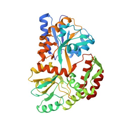 | |
UniProt | |||||
Find proteins for P19576 (Salmonella typhimurium (strain LT2 / SGSC1412 / ATCC 700720)) Explore P19576 Go to UniProtKB: P19576 | |||||
Entity Groups | |||||
| Sequence Clusters | 30% Identity50% Identity70% Identity90% Identity95% Identity100% Identity | ||||
| UniProt Group | P19576 | ||||
Sequence AnnotationsExpand | |||||
| |||||
| Ligands 6 Unique | |||||
|---|---|---|---|---|---|
| ID | Chains | Name / Formula / InChI Key | 2D Diagram | 3D Interactions | |
| ASO Query on ASO | K [auth A], L [auth A], M [auth A] | 1,5-anhydro-D-glucitol C6 H12 O5 MPCAJMNYNOGXPB-SLPGGIOYSA-N |  | ||
| TRS Query on TRS | N [auth A], O [auth A] | 2-AMINO-2-HYDROXYMETHYL-PROPANE-1,3-DIOL C4 H12 N O3 LENZDBCJOHFCAS-UHFFFAOYSA-O |  | ||
| MPD Query on MPD | C [auth A] | (4S)-2-METHYL-2,4-PENTANEDIOL C6 H14 O2 SVTBMSDMJJWYQN-YFKPBYRVSA-N |  | ||
| GOL Query on GOL | P [auth A], Q [auth A] | GLYCEROL C3 H8 O3 PEDCQBHIVMGVHV-UHFFFAOYSA-N |  | ||
| 2ME Query on 2ME | D [auth A], E [auth A], F [auth A], G [auth A] | METHOXYETHANE C3 H8 O XOBKSJJDNFUZPF-UHFFFAOYSA-N |  | ||
| MG Query on MG | H [auth A], I [auth A], J [auth A] | MAGNESIUM ION Mg JLVVSXFLKOJNIY-UHFFFAOYSA-N |  | ||
Entity ID: 2 | |||||
|---|---|---|---|---|---|
| ID | Chains | Name | Type/Class | 2D Diagram | 3D Interactions |
| PRD_900001 Query on PRD_900001 | B | alpha-maltose | Oligosaccharide / Nutrient |  | |
| Length ( Å ) | Angle ( ˚ ) |
|---|---|
| a = 87.714 | α = 90 |
| b = 89.692 | β = 114.766 |
| c = 64.066 | γ = 90 |
| Software Name | Purpose |
|---|---|
| PHENIX | refinement |
| HKL-2000 | data reduction |
| HKL-2000 | data scaling |
| PHENIX | phasing |
| Funding Organization | Location | Grant Number |
|---|---|---|
| National Natural Science Foundation of China | China | 31200556 |
| National Natural Science Foundation of China | China | 21272031 |
RCSB PDB is hosted by

RCSB PDB is a member of the


