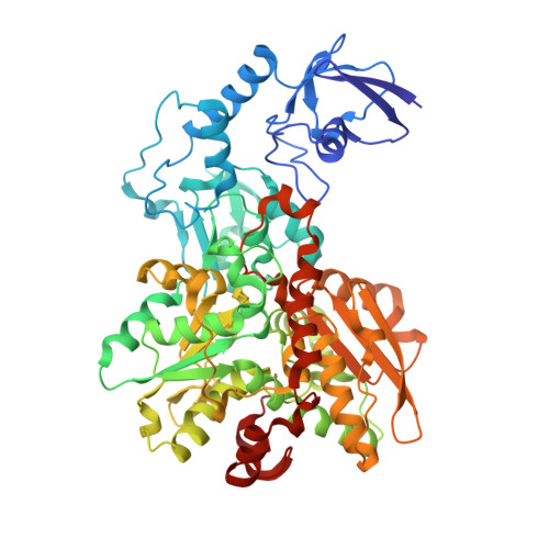
6NAC

Crystal structure of [FeFe]-hydrogenase I (CpI) solved with single pulse free electron laser data


Experimental Data Snapshot

  • Method: X-RAY DIFFRACTION
  • Resolution: 1.55 Å
  • R-Value Free: 0.285 
  • R-Value Work: 0.239 
  • R-Value Observed: 0.240 

Starting Model: experimental
View more details

wwPDB Validation   3D Report Full Report


Ligand Structure Quality Assessment 


This is version 1.4 of the entry. See complete history


Literature

Tuning Catalytic Bias of Hydrogen Gas Producing Hydrogenases.

Artz, J.H.Zadvornyy, O.A.Mulder, D.W.Keable, S.M.Cohen, A.E.Ratzloff, M.W.Williams, S.G.Ginovska, B.Kumar, N.Song, J.McPhillips, S.E.Davidson, C.M.Lyubimov, A.Y.Pence, N.Schut, G.J.Jones, A.K.Soltis, S.M.Adams, M.W.W.Raugei, S.King, P.W.Peters, J.W.

(2020) J Am Chem Soc 142: 1227-1235

  • DOI: https://doi.org/10.1021/jacs.9b08756
  • Primary Citation of Related Structures:  
    6N59, 6N6P, 6NAC

  • PubMed Abstract: 

    Hydrogenases display a wide range of catalytic rates and biases in reversible hydrogen gas oxidation catalysis. The interactions of the iron-sulfur-containing catalytic site with the local protein environment are thought to contribute to differences in catalytic reactivity, but this has not been demonstrated. The microbe Clostridium pasteurianum produces three [FeFe]-hydrogenases that differ in "catalytic bias" by exerting a disproportionate rate acceleration in one direction or the other that spans a remarkable 6 orders of magnitude. The combination of high-resolution structural work, biochemical analyses, and computational modeling indicates that protein secondary interactions directly influence the relative stabilization/destabilization of different oxidation states of the active site metal cluster. This selective stabilization or destabilization of oxidation states can preferentially promote hydrogen oxidation or proton reduction and represents a simple yet elegant model by which a protein catalytic site can confer catalytic bias.


  • Organizational Affiliation

    Institute of Biological Chemistry , Washington State University , Pullman , Washington 99164 , United States.


Macromolecules
Find similar proteins by:  (by identity cutoff)  |  3D Structure
Entity ID: 1
MoleculeChains Sequence LengthOrganismDetailsImage
Iron hydrogenase 1574Clostridium pasteurianumMutation(s): 0 
EC: 1.12.7.2
UniProt
Find proteins for P29166 (Clostridium pasteurianum)
Go to UniProtKB:  P29166
Sequence Annotations
Expand
  • Reference Sequence
Small Molecules
Ligands 4 Unique
IDChains Name / Formula / InChI Key2D Diagram3D Interactions
402
Query on 402

Download Ideal Coordinates CCD File 
B [auth A]dicarbonyl[bis(cyanide-kappaC)]-mu-(iminodimethanethiolatato-1kappaS:2kappaS)-mu-(oxomethylidene)diiron(2+)
C7 H5 Fe2 N3 O3 S2
LJPDYWPSPOWMIB-UHFFFAOYSA-N
SF4
Query on SF4

Download Ideal Coordinates CCD File 
C [auth A],
D [auth A],
E [auth A],
F [auth A]
IRON/SULFUR CLUSTER
Fe4 S4
LJBDFODJNLIPKO-UHFFFAOYSA-N
FES
Query on FES

Download Ideal Coordinates CCD File 
G [auth A]FE2/S2 (INORGANIC) CLUSTER
Fe2 S2
NIXDOXVAJZFRNF-UHFFFAOYSA-N
GOL
Query on GOL

Download Ideal Coordinates CCD File 
H [auth A],
I [auth A]
GLYCEROL
C3 H8 O3
PEDCQBHIVMGVHV-UHFFFAOYSA-N
Experimental Data & Validation

Experimental Data

  • Method: X-RAY DIFFRACTION
  • Resolution: 1.55 Å
  • R-Value Free: 0.285 
  • R-Value Work: 0.239 
  • R-Value Observed: 0.240 
  • Space Group: P 42 21 2
Unit Cell:
Length ( Å )Angle ( ˚ )
a = 111.165α = 90
b = 111.165β = 90
c = 104.007γ = 90
Software Package:
Software NamePurpose
PHENIXrefinement
cctbx.xfeldata reduction
cctbx.primedata scaling
MOLREPphasing

Structure Validation

View Full Validation Report


Ligand Structure Quality Assessment 


Entry History & Funding Information

Deposition Data


Funding OrganizationLocationGrant Number
Department of Energy (DOE, United States)United StatesDE-SC0012518
Department of Energy (DOE, United States)United StatesDE-AC02-76SF00515
National Institutes of Health/National Human Genome Research Institute (NIH/NHGRI)United StatesP41GM103393

Revision History  (Full details and data files)

  • Version 1.0: 2019-12-11
    Type: Initial release
  • Version 1.1: 2019-12-25
    Changes: Database references
  • Version 1.2: 2020-01-22
    Changes: Database references
  • Version 1.3: 2020-02-05
    Changes: Database references
  • Version 1.4: 2023-10-11
    Changes: Data collection, Database references, Derived calculations, Refinement description