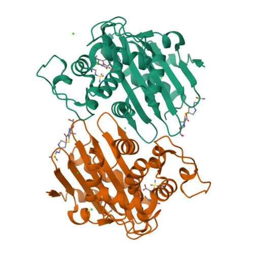
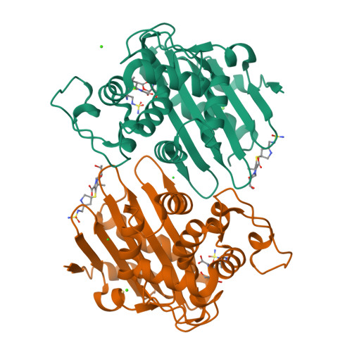
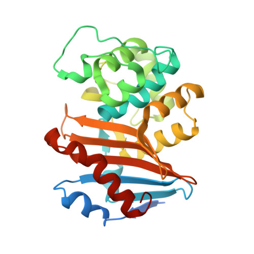
6P9C

OXA-48 carbapanemase, doripenem complex


Experimental Data Snapshot

  • Method: X-RAY DIFFRACTION
  • Resolution: 1.90 Å
  • R-Value Free: 0.241 
  • R-Value Work: 0.194 
  • R-Value Observed: 0.197 

wwPDB Validation   3D Report Full Report


Ligand Structure Quality Assessment 

Created with Raphaël 2.3.0Worse 01 BetterLigand structure goodness of fit to experimental dataBest fitted 4J6Click on this verticalbar to view details

This is version 1.2 of the entry. See complete history


Literature

Structural Insights into the Mechanism of Carbapenemase Activity of the OXA-48 beta-Lactamase.

Smith, C.A.Stewart, N.K.Toth, M.Vakulenko, S.B.

(2019) Antimicrob Agents Chemother 63

  • DOI: https://doi.org/10.1128/AAC.01202-19
  • Primary Citation of Related Structures:  
    6P96, 6P97, 6P98, 6P99, 6P9C

  • PubMed Abstract: 

    Carbapenem-hydrolyzing class D carbapenemases (CHDLs) are enzymes that produce resistance to the last-resort carbapenem antibiotics, severely compromising the available therapeutic options for the treatment of life-threatening infections. A broad variety of CHDLs, including OXA-23, OXA-24/40, and OXA-58, circulate in Acinetobacter baumannii , while the OXA-48 CHDL is predominant in Enterobacteriaceae Extensive structural studies of A. baumannii enzymes have provided important information regarding their interactions with carbapenems and significantly contributed to the understanding of the mechanism of their carbapenemase activity. However, the interactions between carbapenems and OXA-48 have not yet been elucidated. We determined the X-ray crystal structures of the acyl-enzyme complexes of OXA-48 with four carbapenems, imipenem, meropenem, ertapenem, and doripenem, and compared them with those of known carbapenem complexes of A. baumannii CHDLs. In the A. baumannii enzymes, acylation by carbapenems triggers significant displacement of one of two conserved hydrophobic surface residues, resulting in the formation of a channel for entry of the deacylating water into the active site. We show that such a channel preexists in apo-OXA-48 and that only minor displacement of the conserved hydrophobic surface residues occurs upon the formation of OXA-48 acyl-enzyme intermediates. We also demonstrate that the extensive hydrophobic interactions that occur between a conserved hydrophobic bridge of the A. baumannii CHDLs and the carbapenem tails are lost in OXA-48 in the absence of an equivalent bridge structure. These data highlight significant differences between the interactions of carbapenems with OXA-48 and those with A. baumannii enzymes and provide important insights into the mechanism of carbapenemase activity of the major Enterobacteriaceae CHDL, OXA-48.


  • Organizational Affiliation

    Stanford Synchrotron Radiation Lightsource, Stanford University, Menlo Park, California, USA csmith@slac.stanford.edu svakulen@nd.edu.


Macromolecules
Find similar proteins by:  (by identity cutoff)  |  3D Structure
Entity ID: 1
MoleculeChains Sequence LengthOrganismDetailsImage
Beta-lactamase
A, B
265Klebsiella pneumoniaeMutation(s): 0 
Gene Names: bla OXA-48blaOXA-48KPE71T_00045
EC: 3.5.2.6
UniProt
Find proteins for Q6XEC0 (Klebsiella pneumoniae)
Explore Q6XEC0 
Go to UniProtKB:  Q6XEC0
Entity Groups  
Sequence Clusters30% Identity50% Identity70% Identity90% Identity95% Identity100% Identity
UniProt GroupQ6XEC0
Sequence Annotations
Expand
  • Reference Sequence
Small Molecules
Ligands 5 Unique
IDChains Name / Formula / InChI Key2D Diagram3D Interactions
4J6 (Subject of Investigation/LOI)
Query on 4J6

Download Ideal Coordinates CCD File 
C [auth A],
J [auth B]
(4R,5S)-5-[(2S,3R)-3-hydroxy-1-oxobutan-2-yl]-4-methyl-3-({(3S,5S)-5-[(sulfamoylamino)methyl]pyrrolidin-3-yl}sulfanyl)-4,5-dihydro-1H-pyrrole-2-carboxylic acid
C15 H26 N4 O6 S2
QJKQOMNNBOTMCR-PQTSNVLCSA-N
O6P
Query on O6P

Download Ideal Coordinates CCD File 
D [auth A],
K [auth B]
Doripenem
C15 H24 N4 O6 S2
AVAACINZEOAHHE-VFZPANTDSA-N
CD
Query on CD

Download Ideal Coordinates CCD File 
I [auth A],
O [auth B]
CADMIUM ION
Cd
WLZRMCYVCSSEQC-UHFFFAOYSA-N
CA
Query on CA

Download Ideal Coordinates CCD File 
G [auth A],
H [auth A],
N [auth B]
CALCIUM ION
Ca
BHPQYMZQTOCNFJ-UHFFFAOYSA-N
CL
Query on CL

Download Ideal Coordinates CCD File 
E [auth A],
F [auth A],
L [auth B],
M [auth B]
CHLORIDE ION
Cl
VEXZGXHMUGYJMC-UHFFFAOYSA-M
Experimental Data & Validation

Experimental Data

  • Method: X-RAY DIFFRACTION
  • Resolution: 1.90 Å
  • R-Value Free: 0.241 
  • R-Value Work: 0.194 
  • R-Value Observed: 0.197 
  • Space Group: P 21 21 21
Unit Cell:
Length ( Å )Angle ( ˚ )
a = 72.604α = 90
b = 76.749β = 90
c = 101.188γ = 90
Software Package:
Software NamePurpose
PHENIXrefinement
PDB_EXTRACTdata extraction
XDSdata reduction
Aimlessdata scaling
MOLREPphasing

Structure Validation

View Full Validation Report


Ligand Structure Quality Assessment 

Created with Raphaël 2.3.0Worse 01 BetterLigand structure goodness of fit to experimental dataBest fitted 4J6Click on this verticalbar to view details

Entry History 

Deposition Data

Revision History  (Full details and data files)

  • Version 1.0: 2019-08-07
    Type: Initial release
  • Version 1.1: 2019-10-09
    Changes: Data collection, Database references
  • Version 1.2: 2024-10-23
    Changes: Data collection, Database references, Derived calculations, Structure summary