6S4F

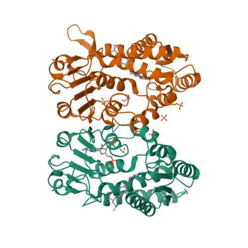
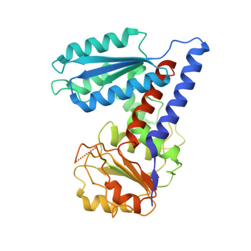
Structure of human MTHFD2 in complex with TH9619


Experimental Data Snapshot

  • Method: X-RAY DIFFRACTION
  • Resolution: 2.20 Å
  • R-Value Free: 0.225 
  • R-Value Work: 0.184 
  • R-Value Observed: 0.186 

wwPDB Validation   3D Report Full Report


Ligand Structure Quality Assessment 


This is version 2.1 of the entry. See complete history


Literature

Pharmacological targeting of MTHFD2 suppresses acute myeloid leukemia by inducing thymidine depletion and replication stress.

Bonagas, N.Gustafsson, N.M.S.Henriksson, M.Marttila, P.Gustafsson, R.Wiita, E.Borhade, S.Green, A.C.Vallin, K.S.A.Sarno, A.Svensson, R.Gokturk, C.Pham, T.Jemth, A.S.Loseva, O.Cookson, V.Kiweler, N.Sandberg, L.Rasti, A.Unterlass, J.E.Haraldsson, M.Andersson, Y.Scaletti, E.R.Bengtsson, C.Paulin, C.B.J.Sanjiv, K.Abdurakhmanov, E.Pudelko, L.Kunz, B.Desroses, M.Iliev, P.Farnegardh, K.Kramer, A.Garg, N.Michel, M.Haggblad, S.Jarvius, M.Kalderen, C.Jensen, A.B.Almlof, I.Karsten, S.Zhang, S.M.Haggblad, M.Eriksson, A.Liu, J.Glinghammar, B.Nekhotiaeva, N.Klingegard, F.Koolmeister, T.Martens, U.Llona-Minguez, S.Moulson, R.Nordstrom, H.Parrow, V.Dahllund, L.Sjoberg, B.Vargas, I.L.Vo, D.D.Wannberg, J.Knapp, S.Krokan, H.E.Arvidsson, P.I.Scobie, M.Meiser, J.Stenmark, P.Berglund, U.W.Homan, E.J.Helleday, T.

(2022) Nat Cancer 3: 156-172

  • DOI: https://doi.org/10.1038/s43018-022-00331-y
  • Primary Citation of Related Structures:  
    6S4A, 6S4E, 6S4F

  • PubMed Abstract: 

    The folate metabolism enzyme MTHFD2 (methylenetetrahydrofolate dehydrogenase/cyclohydrolase) is consistently overexpressed in cancer but its roles are not fully characterized, and current candidate inhibitors have limited potency for clinical development. In the present study, we demonstrate a role for MTHFD2 in DNA replication and genomic stability in cancer cells, and perform a drug screen to identify potent and selective nanomolar MTHFD2 inhibitors; protein cocrystal structures demonstrated binding to the active site of MTHFD2 and target engagement. MTHFD2 inhibitors reduced replication fork speed and induced replication stress followed by S-phase arrest and apoptosis of acute myeloid leukemia cells in vitro and in vivo, with a therapeutic window spanning four orders of magnitude compared with nontumorigenic cells. Mechanistically, MTHFD2 inhibitors prevented thymidine production leading to misincorporation of uracil into DNA and replication stress. Overall, these results demonstrate a functional link between MTHFD2-dependent cancer metabolism and replication stress that can be exploited therapeutically with this new class of inhibitors.


  • Organizational Affiliation

    Science for Life Laboratory, Department of Oncology-Pathology, Karolinska Institutet, Solna, Sweden.


Macromolecules
Find similar proteins by:  (by identity cutoff)  |  3D Structure
Entity ID: 1
MoleculeChains Sequence LengthOrganismDetailsImage
Bifunctional methylenetetrahydrofolate dehydrogenase/cyclohydrolase, mitochondrial
A, B
316Homo sapiensMutation(s): 0 
Gene Names: MTHFD2NMDMC
EC: 1.5.1.15 (PDB Primary Data), 3.5.4.9 (PDB Primary Data)
UniProt & NIH Common Fund Data Resources
Find proteins for P13995 (Homo sapiens)
Explore P13995 
Go to UniProtKB:  P13995
PHAROS:  P13995
GTEx:  ENSG00000065911 
Entity Groups  
Sequence Clusters30% Identity50% Identity70% Identity90% Identity95% Identity100% Identity
UniProt GroupP13995
Sequence Annotations
Expand
  • Reference Sequence
Small Molecules
Ligands 4 Unique
IDChains Name / Formula / InChI Key2D Diagram3D Interactions
KUN (Subject of Investigation/LOI)
Query on KUN

Download Ideal Coordinates CCD File 
C [auth A],
K [auth B]
(E,4S)-4-[[5-[2-[2,6-bis(azanyl)-4-oxidanylidene-1H-pyrimidin-5-yl]ethanoylamino]-3-fluoranyl-pyridin-2-yl]carbonylamino]pent-2-enedioic acid
C17 H18 F N7 O7
ALGPVOJFIGPPPX-VIFPVBQESA-N
ADP
Query on ADP

Download Ideal Coordinates CCD File 
I [auth A]ADENOSINE-5'-DIPHOSPHATE
C10 H15 N5 O10 P2
XTWYTFMLZFPYCI-KQYNXXCUSA-N
PO4
Query on PO4

Download Ideal Coordinates CCD File 
D [auth A]
E [auth A]
F [auth A]
G [auth A]
H [auth A]
D [auth A],
E [auth A],
F [auth A],
G [auth A],
H [auth A],
L [auth B],
M [auth B],
N [auth B]
PHOSPHATE ION
O4 P
NBIIXXVUZAFLBC-UHFFFAOYSA-K
GOL
Query on GOL

Download Ideal Coordinates CCD File 
J [auth A],
O [auth B]
GLYCEROL
C3 H8 O3
PEDCQBHIVMGVHV-UHFFFAOYSA-N
Experimental Data & Validation

Experimental Data

  • Method: X-RAY DIFFRACTION
  • Resolution: 2.20 Å
  • R-Value Free: 0.225 
  • R-Value Work: 0.184 
  • R-Value Observed: 0.186 
  • Space Group: P 65
Unit Cell:
Length ( Å )Angle ( ˚ )
a = 116.895α = 90
b = 116.895β = 90
c = 112.965γ = 120
Software Package:
Software NamePurpose
REFMACrefinement
DIALSdata reduction
Aimlessdata scaling
PHASERphasing

Structure Validation

View Full Validation Report


Ligand Structure Quality Assessment 


Entry History 

Revision History  (Full details and data files)

  • Version 1.0: 2021-07-07
    Type: Initial release
  • Version 1.1: 2022-05-11
    Changes: Advisory, Data collection, Database references, Derived calculations, Structure summary
  • Version 2.0: 2023-04-12
    Changes: Non-polymer description, Structure summary
  • Version 2.1: 2024-06-19
    Changes: Data collection