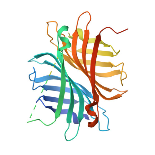
6S50

scdSav(SARK)mv2 - Engineering Single-Chain Dimeric Streptavidin as Host for Artificial Metalloenzymes


Experimental Data Snapshot

  • Method: X-RAY DIFFRACTION
  • Resolution: 2.00 Å
  • R-Value Free: 0.208 
  • R-Value Work: 0.185 
  • R-Value Observed: 0.186 

Starting Model: experimental
View more details

wwPDB Validation   3D Report Full Report


Ligand Structure Quality Assessment 


This is version 1.3 of the entry. See complete history


Literature

Breaking Symmetry: Engineering Single-Chain Dimeric Streptavidin as Host for Artificial Metalloenzymes.

Wu, S.Zhou, Y.Rebelein, J.G.Kuhn, M.Mallin, H.Zhao, J.Igareta, N.V.Ward, T.R.

(2019) J Am Chem Soc 141: 15869-15878

  • DOI: https://doi.org/10.1021/jacs.9b06923
  • Primary Citation of Related Structures:  
    6S4Q, 6S50

  • PubMed Abstract: 

    The biotin-streptavidin technology has been extensively exploited to engineer artificial metalloenzymes (ArMs) that catalyze a dozen different reactions. Despite its versatility, the homotetrameric nature of streptavidin (Sav) and the noncooperative binding of biotinylated cofactors impose two limitations on the genetic optimization of ArMs: (i) point mutations are reflected in all four subunits of Sav, and (ii) the noncooperative binding of biotinylated cofactors to Sav may lead to an erosion in the catalytic performance, depending on the cofactor:biotin-binding site ratio. To address these challenges, we report on our efforts to engineer a (monovalent) single-chain dimeric streptavidin (scdSav) as scaffold for Sav-based ArMs. The versatility of scdSav as host protein is highlighted for the asymmetric transfer hydrogenation of prochiral imines using [Cp*Ir(biot- p -L)Cl] as cofactor. By capitalizing on a more precise genetic fine-tuning of the biotin-binding vestibule, unrivaled levels of activity and selectivity were achieved for the reduction of challenging prochiral imines. Comparison of the saturation kinetic data and X-ray structures of [Cp*Ir(biot- p -L)Cl]·scdSav with a structurally related [Cp*Ir(biot- p -L)Cl]·monovalent scdSav highlights the advantages of the presence of a single biotinylated cofactor precisely localized within the biotin-binding vestibule of the monovalent scdSav. The practicality of scdSav-based ArMs was illustrated for the reduction of the salsolidine precursor (500 mM) to afford ( R )-salsolidine in 90% ee and >17 000 TONs. Monovalent scdSav thus provides a versatile scaffold to evolve more efficient ArMs for in vivo catalysis and large-scale applications.


  • Organizational Affiliation

    Department of Chemistry , University of Basel , BPR 1096, Mattenstrasse 24a , CH-4058 Basel , Switzerland.


Macromolecules
Find similar proteins by:  (by identity cutoff)  |  3D Structure
Entity ID: 1
MoleculeChains Sequence LengthOrganismDetailsImage
Streptavidin
A, B
343Streptomyces avidiniiMutation(s): 0 
UniProt
Find proteins for P22629 (Streptomyces avidinii)
Explore P22629 
Go to UniProtKB:  P22629
Entity Groups  
Sequence Clusters30% Identity50% Identity70% Identity90% Identity95% Identity100% Identity
UniProt GroupP22629
Sequence Annotations
Expand
  • Reference Sequence
Small Molecules
Ligands 3 Unique
IDChains Name / Formula / InChI Key2D Diagram3D Interactions
4IR
Query on 4IR

Download Ideal Coordinates CCD File 
C [auth A],
D [auth A],
I [auth B],
J [auth B]
{N-(4-{[2-(amino-kappaN)ethyl]sulfamoyl-kappaN}phenyl)-5-[(3aS,4S,6aR)-2-oxohexahydro-1H-thieno[3,4-d]imidazol-4-yl]pentanamide}(chloro)[(1,2,3,4,5-eta)-1,2,3,4,5-pentamethylcyclopentadienyl]iridium(III)
C28 H45 Cl Ir N5 O4 S2
OPJZDGOSRZOXTM-DYKJULMGSA-M
SO4
Query on SO4

Download Ideal Coordinates CCD File 
H [auth A]SULFATE ION
O4 S
QAOWNCQODCNURD-UHFFFAOYSA-L
GOL
Query on GOL

Download Ideal Coordinates CCD File 
E [auth A]
F [auth A]
G [auth A]
K [auth B]
L [auth B]
E [auth A],
F [auth A],
G [auth A],
K [auth B],
L [auth B],
M [auth B]
GLYCEROL
C3 H8 O3
PEDCQBHIVMGVHV-UHFFFAOYSA-N
Experimental Data & Validation

Experimental Data

  • Method: X-RAY DIFFRACTION
  • Resolution: 2.00 Å
  • R-Value Free: 0.208 
  • R-Value Work: 0.185 
  • R-Value Observed: 0.186 
  • Space Group: C 1 2 1
Unit Cell:
Length ( Å )Angle ( ˚ )
a = 183.879α = 90
b = 57.771β = 108.33
c = 57.645γ = 90
Software Package:
Software NamePurpose
PHENIXrefinement
XDSdata reduction
Aimlessdata scaling
PHASERphasing

Structure Validation

View Full Validation Report


Ligand Structure Quality Assessment 


Entry History & Funding Information

Deposition Data


Funding OrganizationLocationGrant Number
Swiss National Science FoundationSwitzerland200020_182046
European Research CouncilDrEAM
Swiss National Science FoundationSwitzerlandNCCR MSE
European Molecular Biology OrganizationGermanyALTF 194-2017

Revision History  (Full details and data files)

  • Version 1.0: 2019-09-25
    Type: Initial release
  • Version 1.1: 2019-10-16
    Changes: Data collection, Database references
  • Version 1.2: 2024-01-24
    Changes: Data collection, Database references, Refinement description
  • Version 1.3: 2024-11-20
    Changes: Structure summary