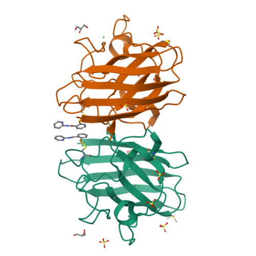
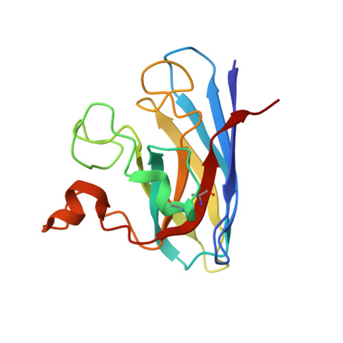
6SPJ

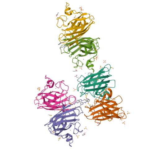
A4V MUTANT OF HUMAN SOD1 WITH EBSELEN DERIVATIVE 1


Experimental Data Snapshot

  • Method: X-RAY DIFFRACTION
  • Resolution: 1.97 Å
  • R-Value Free: 0.225 
  • R-Value Work: 0.182 
  • R-Value Observed: 0.185 

Starting Model: experimental
View more details

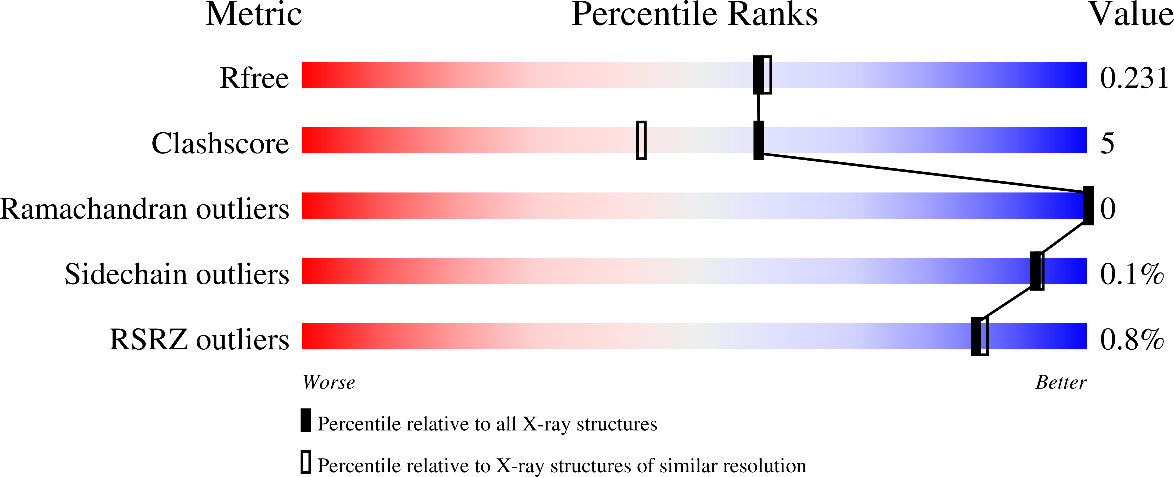
wwPDB Validation   3D Report Full Report


Ligand Structure Quality Assessment 


This is version 1.2 of the entry. See complete history


Literature

Ebselen as template for stabilization of A4V mutant dimer for motor neuron disease therapy.

Chantadul, V.Wright, G.S.A.Amporndanai, K.Shahid, M.Antonyuk, S.V.Washbourn, G.Rogers, M.Roberts, N.Pye, M.O'Neill, P.M.Hasnain, S.S.

(2020) Commun Biol 3: 97-97

  • DOI: https://doi.org/10.1038/s42003-020-0826-3
  • Primary Citation of Related Structures:  
    6SPA, 6SPH, 6SPI, 6SPJ, 6SPK

  • PubMed Abstract: 

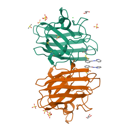
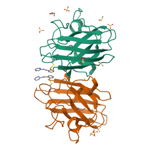
    Mutations to the gene encoding superoxide dismutase-1 (SOD1) were the first genetic elements discovered that cause motor neuron disease (MND). These mutations result in compromised SOD1 dimer stability, with one of the severest and most common mutations Ala4Val (A4V) displaying a propensity to monomerise and aggregate leading to neuronal death. We show that the clinically used ebselen and related analogues promote thermal stability of A4V SOD1 when binding to Cys111 only. We have developed a A4V SOD1 differential scanning fluorescence-based assay on a C6S mutation background that is effective in assessing suitability of compounds. Crystallographic data show that the selenium atom of these compounds binds covalently to A4V SOD1 at Cys111 at the dimer interface, resulting in stabilisation. This together with chemical amenability for hit expansion of ebselen and its on-target SOD1 pharmacological chaperone activity holds remarkable promise for structure-based therapeutics for MND using ebselen as a template.


  • Organizational Affiliation

    Faculty of Health and Life Sciences, Molecular Biophysics Group, Institute of Integrative Biology, University of Liverpool, Liverpool, L69 7ZB, UK.


Macromolecules
Find similar proteins by:  (by identity cutoff)  |  3D Structure
Entity ID: 1
MoleculeChains Sequence LengthOrganismDetailsImage
Superoxide dismutase [Cu-Zn]153Homo sapiensMutation(s): 1 
Gene Names: SOD1
EC: 1.15.1.1
UniProt & NIH Common Fund Data Resources
Find proteins for P00441 (Homo sapiens)
Explore P00441 
Go to UniProtKB:  P00441
PHAROS:  P00441
GTEx:  ENSG00000142168 
Entity Groups  
Sequence Clusters30% Identity50% Identity70% Identity90% Identity95% Identity100% Identity
UniProt GroupP00441
Sequence Annotations
Expand
  • Reference Sequence
Small Molecules
Ligands 6 Unique
IDChains Name / Formula / InChI Key2D Diagram3D Interactions
LR2 (Subject of Investigation/LOI)
Query on LR2

Download Ideal Coordinates CCD File 
CB [auth I]
H [auth A]
HA [auth E]
NB [auth J]
U [auth B]
CB [auth I],
H [auth A],
HA [auth E],
NB [auth J],
U [auth B],
UA [auth F]
~{N}-pyridin-2-yl-2-selanyl-benzamide
C12 H10 N2 O Se
IIISTDSZDJBBTA-UHFFFAOYSA-N
SO4
Query on SO4

Download Ideal Coordinates CCD File 
AA [auth B]
BA [auth B]
CA [auth B]
EB [auth I]
FB [auth I]
AA [auth B],
BA [auth B],
CA [auth B],
EB [auth I],
FB [auth I],
GB [auth I],
HB [auth I],
IB [auth I],
J [auth A],
JA [auth E],
K [auth A],
KA [auth E],
L [auth A],
LA [auth E],
M [auth A],
MA [auth E],
N [auth A],
O [auth A],
PB [auth J],
QB [auth J],
RB [auth J],
SB [auth J],
TB [auth J],
W [auth B],
WA [auth F],
X [auth B],
XA [auth F],
Y [auth B],
YA [auth F],
Z [auth B]
SULFATE ION
O4 S
QAOWNCQODCNURD-UHFFFAOYSA-L
GOL
Query on GOL

Download Ideal Coordinates CCD File 
DA [auth B]
JB [auth I]
NA [auth E]
P [auth A]
UB [auth J]
DA [auth B],
JB [auth I],
NA [auth E],
P [auth A],
UB [auth J],
ZA [auth F]
GLYCEROL
C3 H8 O3
PEDCQBHIVMGVHV-UHFFFAOYSA-N
DMS
Query on DMS

Download Ideal Coordinates CCD File 
EA [auth B]
KB [auth I]
OA [auth E]
PA [auth E]
Q [auth A]
EA [auth B],
KB [auth I],
OA [auth E],
PA [auth E],
Q [auth A],
QA [auth E],
R [auth A],
RA [auth E],
VB [auth J]
DIMETHYL SULFOXIDE
C2 H6 O S
IAZDPXIOMUYVGZ-UHFFFAOYSA-N
ZN
Query on ZN

Download Ideal Coordinates CCD File 
BB [auth I]
DB [auth I]
G [auth A]
GA [auth E]
I [auth A]
BB [auth I],
DB [auth I],
G [auth A],
GA [auth E],
I [auth A],
IA [auth E],
MB [auth J],
OB [auth J],
T [auth B],
TA [auth F],
V [auth B],
VA [auth F]
ZINC ION
Zn
PTFCDOFLOPIGGS-UHFFFAOYSA-N
CL
Query on CL

Download Ideal Coordinates CCD File 
AB [auth F]
FA [auth B]
LB [auth I]
S [auth A]
SA [auth E]
AB [auth F],
FA [auth B],
LB [auth I],
S [auth A],
SA [auth E],
WB [auth J]
CHLORIDE ION
Cl
VEXZGXHMUGYJMC-UHFFFAOYSA-M
Experimental Data & Validation

Experimental Data

  • Method: X-RAY DIFFRACTION
  • Resolution: 1.97 Å
  • R-Value Free: 0.225 
  • R-Value Work: 0.182 
  • R-Value Observed: 0.185 
  • Space Group: C 1 2 1
Unit Cell:
Length ( Å )Angle ( ˚ )
a = 112.506α = 90
b = 194.658β = 96.97
c = 75.169γ = 90
Software Package:
Software NamePurpose
REFMACrefinement
Aimlessdata scaling
PDB_EXTRACTdata extraction
DIALSdata reduction
MOLREPphasing

Structure Validation

View Full Validation Report


Ligand Structure Quality Assessment 


Entry History 

Deposition Data

Revision History  (Full details and data files)

  • Version 1.0: 2020-03-18
    Type: Initial release
  • Version 1.1: 2024-01-24
    Changes: Data collection, Database references, Derived calculations, Refinement description
  • Version 1.2: 2024-11-20
    Changes: Structure summary