6SPR | pdb_00006spr

Structure of the Escherichia coli methionyl-tRNA synthetase variant VI298 complexed with beta-methionine


Experimental Data Snapshot

  • Method: X-RAY DIFFRACTION
  • Resolution: 1.48 Å
  • R-Value Free: 
    0.168 (Depositor), 0.170 (DCC) 
  • R-Value Work: 
    0.131 (Depositor), 0.130 (DCC) 
  • R-Value Observed: 
    0.133 (Depositor) 

Starting Model: experimental
View more details

wwPDB Validation   3D Report Full Report


Ligand Structure Quality Assessment 

Created with Raphaël 2.3.0Worse 01 BetterLigand structure goodness of fit to experimental dataBest fitted B3MClick on this verticalbar to view details

This is version 2.1 of the entry. See complete history


Literature

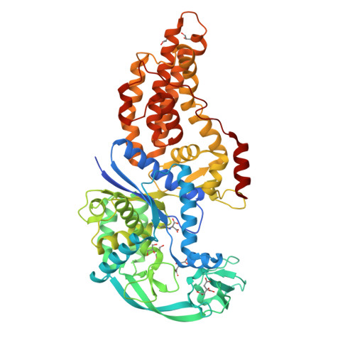
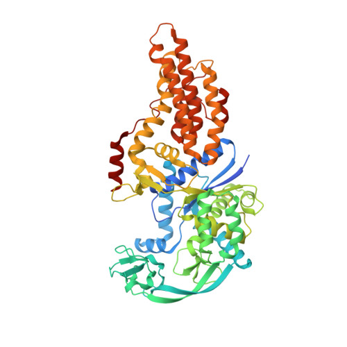
Use of beta3-methionine as an amino acid substrate of Escherichia coli methionyl-tRNA synthetase.

Nigro, G.Bourcier, S.Lazennec-Schurdevin, C.Schmitt, E.Marliere, P.Mechulam, Y.

(2020) J Struct Biol 209: 107435-107435

  • DOI: https://doi.org/10.1016/j.jsb.2019.107435
  • Primary Citation of Related Structures:  
    6SPN, 6SPO, 6SPP, 6SPQ, 6SPR

  • PubMed Abstract: 

    Polypeptides containing β-amino acids are attractive tools for the design of novel proteins having unique properties of medical or industrial interest. Incorporation of β-amino acids in vivo requires the development of efficient aminoacyl-tRNA synthetases specific of these non-canonical amino acids. Here, we have performed a detailed structural and biochemical study of the recognition and use of β 3 -Met by Escherichia coli methionyl-tRNA synthetase (MetRS). We show that MetRS binds β 3 -Met with a 24-fold lower affinity but catalyzes the esterification of the non-canonical amino acid onto tRNA with a rate lowered by three orders of magnitude. Accurate measurements of the catalytic parameters required careful consideration of the presence of contaminating α-Met in β 3 -Met commercial samples. The 1.45 Å crystal structure of the MetRS: β 3 -Met complex shows that β 3 -Met binds the enzyme essentially like α-Met, but the carboxylate moiety is mobile and not adequately positioned to react with ATP for aminoacyl adenylate formation. This study provides structural and biochemical bases for engineering MetRS with improved β 3 -Met aminoacylation capabilities.


  • Organizational Affiliation

    Laboratoire de Biochimie, BIOC, Ecole polytechnique, CNRS, Institut Polytechnique de Paris, 91128 Palaiseau Cedex, France.


Macromolecules
Find similar proteins by:  (by identity cutoff)  |  3D Structure
Entity ID: 1
MoleculeChains Sequence LengthOrganismDetailsImage
Methionine--tRNA ligase568Escherichia coliMutation(s): 1 
Gene Names: metGb2114JW2101
EC: 6.1.1.10
UniProt
Find proteins for P00959 (Escherichia coli (strain K12))
Explore P00959 
Go to UniProtKB:  P00959
Entity Groups  
Sequence Clusters30% Identity50% Identity70% Identity90% Identity95% Identity100% Identity
UniProt GroupP00959
Sequence Annotations
Expand
  • Reference Sequence
Small Molecules
Ligands 4 Unique
IDChains Name / Formula / InChI Key2D Diagram3D Interactions
CIT
Query on CIT

Download Ideal Coordinates CCD File 
D [auth A],
E [auth A]
CITRIC ACID
C6 H8 O7
KRKNYBCHXYNGOX-UHFFFAOYSA-N
B3M (Subject of Investigation/LOI)
Query on B3M

Download Ideal Coordinates CCD File 
C [auth A](3R)-3-amino-5-(methylsulfanyl)pentanoic acid
C6 H13 N O2 S
QWVNCDVONVDGDV-YFKPBYRVSA-N
GOL
Query on GOL

Download Ideal Coordinates CCD File 
F [auth A],
G [auth A],
H [auth A],
I [auth A]
GLYCEROL
C3 H8 O3
PEDCQBHIVMGVHV-UHFFFAOYSA-N
ZN
Query on ZN

Download Ideal Coordinates CCD File 
B [auth A]ZINC ION
Zn
PTFCDOFLOPIGGS-UHFFFAOYSA-N
Experimental Data & Validation

Experimental Data

  • Method: X-RAY DIFFRACTION
  • Resolution: 1.48 Å
  • R-Value Free:  0.168 (Depositor), 0.170 (DCC) 
  • R-Value Work:  0.131 (Depositor), 0.130 (DCC) 
  • R-Value Observed: 0.133 (Depositor) 
Space Group: P 1 21 1
Unit Cell:
Length ( Å )Angle ( ˚ )
a = 78.31α = 90
b = 45.11β = 107.39
c = 86.21γ = 90
Software Package:
Software NamePurpose
PHENIXrefinement
PHENIXrefinement
XDSdata reduction
XDSdata scaling
PHENIXphasing

Structure Validation

View Full Validation Report


Ligand Structure Quality Assessment 

Created with Raphaël 2.3.0Worse 01 BetterLigand structure goodness of fit to experimental dataBest fitted B3MClick on this verticalbar to view details

Entry History 

Deposition Data

Revision History  (Full details and data files)

  • Version 1.0: 2020-01-01
    Type: Initial release
  • Version 1.1: 2020-03-11
    Changes: Database references
  • Version 2.0: 2023-11-15
    Changes: Atomic model, Data collection, Database references
  • Version 2.1: 2024-01-24
    Changes: Refinement description