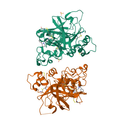
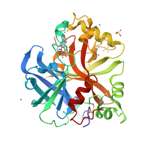
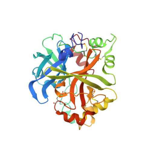
6T8W

Complement factor B in complex with (-)-4-(1-((5,7-Dimethyl-1H-indol-4-yl)methyl)piperidin-2-yl)benzoic acid


Experimental Data Snapshot

  • Method: X-RAY DIFFRACTION
  • Resolution: 1.70 Å
  • R-Value Free: 0.238 
  • R-Value Work: 0.198 

Starting Model: experimental
View more details

wwPDB Validation   3D Report Full Report


Ligand Structure Quality Assessment 


This is version 1.3 of the entry. See complete history


Literature

Discovery of 4-((2S,4S)-4-Ethoxy-1-((5-methoxy-7-methyl-1H-indol-4-yl)methyl)piperidin-2-yl)benzoic Acid (LNP023), a Factor B Inhibitor Specifically Designed To Be Applicable to Treating a Diverse Array of Complement Mediated Diseases.

Mainolfi, N.Ehara, T.Karki, R.G.Anderson, K.Mac Sweeney, A.Liao, S.M.Argikar, U.A.Jendza, K.Zhang, C.Powers, J.Klosowski, D.W.Crowley, M.Kawanami, T.Ding, J.April, M.Forster, C.Serrano-Wu, M.Capparelli, M.Ramqaj, R.Solovay, C.Cumin, F.Smith, T.M.Ferrara, L.Lee, W.Long, D.Prentiss, M.De Erkenez, A.Yang, L.Liu, F.Sellner, H.Sirockin, F.Valeur, E.Erbel, P.Ostermeier, D.Ramage, P.Gerhartz, B.Schubart, A.Flohr, S.Gradoux, N.Feifel, R.Vogg, B.Wiesmann, C.Maibaum, J.Eder, J.Sedrani, R.Harrison, R.A.Mogi, M.Jaffee, B.D.Adams, C.M.

(2020) J Med Chem 63: 5697-5722

  • DOI: https://doi.org/10.1021/acs.jmedchem.9b01870
  • Primary Citation of Related Structures:  
    6T8U, 6T8V, 6T8W

  • PubMed Abstract: 

    The alternative pathway (AP) of the complement system is a key contributor to the pathogenesis of several human diseases including age-related macular degeneration, paroxysmal nocturnal hemoglobinuria (PNH), atypical hemolytic uremic syndrome (aHUS), and various glomerular diseases. The serine protease factor B (FB) is a key node in the AP and is integral to the formation of C3 and C5 convertase. Despite the prominent role of FB in the AP, selective orally bioavailable inhibitors, beyond our own efforts, have not been reported previously. Herein we describe in more detail our efforts to identify FB inhibitors by high-throughput screening (HTS) and leveraging insights from several X-ray cocrystal structures during optimization efforts. This work culminated in the discovery of LNP023 ( 41 ), which is currently being evaluated clinically in several diverse AP mediated indications.


  • Organizational Affiliation

    Novartis Institutes for BioMedical Research, Cambridge, Massachusetts 02139, United States.


Macromolecules
Find similar proteins by:  (by identity cutoff)  |  3D Structure
Entity ID: 1
MoleculeChains Sequence LengthOrganismDetailsImage
Complement factor BA [auth AAA],
B [auth BBB]
291Homo sapiensMutation(s): 0 
Gene Names: CFBBFBFD
EC: 3.4.21.47
UniProt & NIH Common Fund Data Resources
Find proteins for P00751 (Homo sapiens)
Explore P00751 
Go to UniProtKB:  P00751
PHAROS:  P00751
GTEx:  ENSG00000243649 
Entity Groups  
Sequence Clusters30% Identity50% Identity70% Identity90% Identity95% Identity100% Identity
UniProt GroupP00751
Sequence Annotations
Expand
  • Reference Sequence
Small Molecules
Ligands 3 Unique
IDChains Name / Formula / InChI Key2D Diagram3D Interactions
MVW (Subject of Investigation/LOI)
Query on MVW

Download Ideal Coordinates CCD File 
E [auth AAA],
K [auth BBB]
5,7-dimethyl-4-[[(2~{S})-2-phenylpiperidin-1-yl]methyl]-1~{H}-indole
C22 H26 N2
GRJUTCRGMYGLQC-NRFANRHFSA-N
SO4
Query on SO4

Download Ideal Coordinates CCD File 
C [auth AAA],
D [auth AAA],
F [auth BBB],
G [auth BBB],
H [auth BBB]
SULFATE ION
O4 S
QAOWNCQODCNURD-UHFFFAOYSA-L
ZN
Query on ZN

Download Ideal Coordinates CCD File 
I [auth BBB],
J [auth BBB]
ZINC ION
Zn
PTFCDOFLOPIGGS-UHFFFAOYSA-N
Experimental Data & Validation

Experimental Data

  • Method: X-RAY DIFFRACTION
  • Resolution: 1.70 Å
  • R-Value Free: 0.238 
  • R-Value Work: 0.198 
  • Space Group: P 1 21 1
Unit Cell:
Length ( Å )Angle ( ˚ )
a = 54.696α = 90
b = 97.656β = 100.309
c = 61.881γ = 90
Software Package:
Software NamePurpose
REFMACrefinement
XDSdata reduction
XSCALEdata scaling
PHASERphasing

Structure Validation

View Full Validation Report


Ligand Structure Quality Assessment 


Entry History 

Revision History  (Full details and data files)

  • Version 1.0: 2020-03-04
    Type: Initial release
  • Version 1.1: 2020-07-01
    Changes: Database references
  • Version 1.2: 2024-01-24
    Changes: Data collection, Database references, Derived calculations, Refinement description
  • Version 1.3: 2024-11-06
    Changes: Structure summary