6UBO | pdb_00006ubo

Fluorogen Activating Protein Dib1


Experimental Data Snapshot

  • Method: X-RAY DIFFRACTION
  • Resolution: 1.58 Å
  • R-Value Free: 
    0.203 (Depositor), 0.210 (DCC) 
  • R-Value Work: 
    0.163 (Depositor), 0.180 (DCC) 
  • R-Value Observed: 
    0.164 (Depositor) 

Starting Model: experimental
View more details

wwPDB Validation   3D Report Full Report


Ligand Structure Quality Assessment 

Created with Raphaël 2.3.0Worse 01 BetterLigand structure goodness of fit to experimental dataBest fitted Q3JClick on this verticalbar to view details

This is version 1.2 of the entry. See complete history


Literature

Structure-Based Rational Design of Two Enhanced Bacterial Lipocalin Blc Tags for Protein-PAINT Super-resolution Microscopy.

Muslinkina, L.Gavrikov, A.S.Bozhanova, N.G.Mishin, A.S.Baranov, M.S.Meiler, J.Pletneva, N.V.Pletnev, V.Z.Pletnev, S.

(2020) ACS Chem Biol 15: 2456-2465

  • DOI: https://doi.org/10.1021/acschembio.0c00440
  • Primary Citation of Related Structures:  
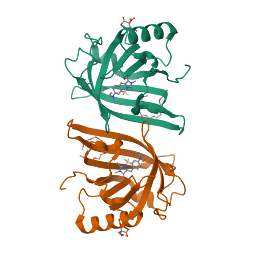
    6UBO

  • PubMed Abstract: 

    Super-resolution fluorescent imaging in living cells remains technically challenging, largely due to the photodecomposition of fluorescent tags. The recently suggested protein-PAINT is the only super-resolution technique available for prolonged imaging of proteins in living cells. It is realized with complexes of fluorogen-activating proteins, expressed as fusions, and solvatochromic synthetic dyes. Once photobleached, the dye in the complex is replaced with a fresh fluorogen available in the sample. With suitable kinetics, this replacement creates fluorescence blinking required for attaining super-resolution and overcomes photobleaching associated with the loss of an irreplaceable fluorophore. Here we report on the rational design of two protein-PAINT tags based on the 1.58 Å crystal structure of the DiB1:M739 complex, an improved green-emitting DiB3/F74V:M739 and a new orange-emitting DiB3/F53L:M739. They outperform previously reported DiB-based tags to become best in class biomarkers for protein-PAINT. The new tags advance protein-PAINT from the proof-of-concept to a reliable tool suitable for prolonged super-resolution imaging of intracellular proteins in fixed and living cells and two-color PAINT-like nanoscopy with a single fluorogen.


  • Organizational Affiliation

    Basic Research Program, Frederick National Laboratory for Cancer Research, Argonne, Illinois 60439, United States.


Macromolecules
Find similar proteins by:  (by identity cutoff)  |  3D Structure
Entity ID: 1
MoleculeChains Sequence LengthOrganismDetailsImage
Outer membrane lipoprotein Blc
A, B
177Escherichia coli K-12Mutation(s): 0 
Gene Names: blcyjeLb4149JW4110
UniProt
Find proteins for P0A901 (Escherichia coli (strain K12))
Explore P0A901 
Go to UniProtKB:  P0A901
Entity Groups  
Sequence Clusters30% Identity50% Identity70% Identity90% Identity95% Identity100% Identity
UniProt GroupP0A901
Sequence Annotations
Expand
  • Reference Sequence
Small Molecules
Ligands 3 Unique
IDChains Name / Formula / InChI Key2D Diagram3D Interactions
P4K
Query on P4K

Download Ideal Coordinates CCD File 
E [auth A],
H [auth B]
polyethylene glycol
C30 H62 O15
WWPGFZAAWXFBTF-UHFFFAOYSA-N
Q3J (Subject of Investigation/LOI)
Query on Q3J

Download Ideal Coordinates CCD File 
C [auth A],
F [auth B]
12-(diethylamino)-2,2-bis(fluoranyl)-4,5-dimethyl-5-aza-3-azonia-2-boranuidatricyclo[7.4.0.0^{3,7}]trideca-1(13),3,7,9,11-pentaen-6-one
C16 H20 B F2 N3 O
OOEJBECJAKZKRT-UHFFFAOYSA-N
CIT
Query on CIT

Download Ideal Coordinates CCD File 
D [auth A],
G [auth B]
CITRIC ACID
C6 H8 O7
KRKNYBCHXYNGOX-UHFFFAOYSA-N
Experimental Data & Validation

Experimental Data

  • Method: X-RAY DIFFRACTION
  • Resolution: 1.58 Å
  • R-Value Free:  0.203 (Depositor), 0.210 (DCC) 
  • R-Value Work:  0.163 (Depositor), 0.180 (DCC) 
  • R-Value Observed: 0.164 (Depositor) 
Space Group: P 43
Unit Cell:
Length ( Å )Angle ( ˚ )
a = 44.843α = 90
b = 44.843β = 90
c = 158.235γ = 90
Software Package:
Software NamePurpose
HKL-2000data scaling
MOLREPphasing
REFMACrefinement
PDB_EXTRACTdata extraction
SERGUIdata collection
HKL-2000data reduction

Structure Validation

View Full Validation Report


Ligand Structure Quality Assessment 

Created with Raphaël 2.3.0Worse 01 BetterLigand structure goodness of fit to experimental dataBest fitted Q3JClick on this verticalbar to view details

Entry History 

Deposition Data

Revision History  (Full details and data files)

  • Version 1.0: 2020-09-16
    Type: Initial release
  • Version 1.1: 2021-03-31
    Changes: Database references
  • Version 1.2: 2023-10-11
    Changes: Data collection, Database references, Refinement description