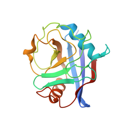
6R8L

Human Cyclophilin D in complex with 1-((1S,9S,10S)-10-Hydroxy-12-oxa-8-aza-tricyclo[7.3.1.02,7]trideca-2,4,6-trien-4-ylmethyl)-3- {2-[(R)-2-(2-methylsulfanyl-phenyl)-pyrrolidin-1-yl]-2-oxo-ethyl}-urea


Experimental Data Snapshot

  • Method: X-RAY DIFFRACTION
  • Resolution: 1.64 Å
  • R-Value Free: 0.203 
  • R-Value Work: 0.173 
  • R-Value Observed: 0.176 

Starting Model: experimental
View more details

wwPDB Validation   3D Report Full Report


Ligand Structure Quality Assessment 


This is version 1.1 of the entry. See complete history


Literature

Discovery of novel Cyclophilin D inhibitors starting from three dimensional fragments with millimolar potencies.

Gradler, U.Schwarz, D.Blaesse, M.Leuthner, B.Johnson, T.L.Bernard, F.Jiang, X.Marx, A.Gilardone, M.Lemoine, H.Roche, D.Jorand-Lebrun, C.

(2019) Bioorg Med Chem Lett 29: 126717-126717

  • DOI: https://doi.org/10.1016/j.bmcl.2019.126717
  • Primary Citation of Related Structures:  
    6R8L, 6R8O, 6R8W, 6R9S, 6R9U, 6R9X, 6RA1

  • PubMed Abstract: 

    Fragment-based screening by SPR enabled the discovery of chemical diverse fragment hits with millimolar binding affinities to the peptidyl-prolyl isomerase Cyclophilin D (CypD). The CypD protein crystal structures of 6 fragment hits provided the basis for subsequent medicinal chemistry optimization by fragment merging and linking yielding three different chemical series with either urea, oxalyl or amide linkers connecting millimolar fragments in the S1' and S2 pockets. We successfully improved the in vitro CypD potencies in the biochemical FP and PPIase assays and in the biophysical SPR binding assay from millimolar towards the low micromolar and submicromolar range by >1000-fold for some fragment derivatives. The initial SAR together with the protein crystal structures of our novel CypD inhibitors provide a suitable basis for further hit-to-lead optimization.


  • Organizational Affiliation

    Merck Healthcare, Merck KGaA, Frankfurter Str. 250, D-64293 Darmstadt, Germany. Electronic address: ulrich.graedler@merckgroup.com.


Macromolecules
Find similar proteins by:  (by identity cutoff)  |  3D Structure
Entity ID: 1
MoleculeChains Sequence LengthOrganismDetailsImage
Peptidyl-prolyl cis-trans isomerase F, mitochondrial164Homo sapiensMutation(s): 1 
Gene Names: PPIFCYP3
EC: 5.2.1.8
UniProt & NIH Common Fund Data Resources
Find proteins for P30405 (Homo sapiens)
Explore P30405 
Go to UniProtKB:  P30405
PHAROS:  P30405
GTEx:  ENSG00000108179 
Entity Groups  
Sequence Clusters30% Identity50% Identity70% Identity90% Identity95% Identity100% Identity
UniProt GroupP30405
Sequence Annotations
Expand
  • Reference Sequence
Small Molecules
Ligands 1 Unique
IDChains Name / Formula / InChI Key2D Diagram3D Interactions
JUZ
Query on JUZ

Download Ideal Coordinates CCD File 
B [auth A]1-[2-[(2~{R})-2-(2-methylsulfanylphenyl)pyrrolidin-1-yl]-2-oxidanylidene-ethyl]-3-[[(1~{S},9~{S},10~{S})-10-oxidanyl-12-oxa-8-azatricyclo[7.3.1.0^{2,7}]trideca-2(7),3,5-trien-4-yl]methyl]urea
C26 H32 N4 O4 S
BICVGZIZTMOLHN-AFXVXQJMSA-N
Experimental Data & Validation

Experimental Data

  • Method: X-RAY DIFFRACTION
  • Resolution: 1.64 Å
  • R-Value Free: 0.203 
  • R-Value Work: 0.173 
  • R-Value Observed: 0.176 
  • Space Group: P 43 21 2
Unit Cell:
Length ( Å )Angle ( ˚ )
a = 57.151α = 90
b = 57.151β = 90
c = 115.386γ = 90
Software Package:
Software NamePurpose
XDSdata reduction
XSCALEdata scaling
REFMACrefinement
MOLREPphasing

Structure Validation

View Full Validation Report


Ligand Structure Quality Assessment 


Entry History 

Deposition Data

  • Released Date: 2019-11-27 
  • Deposition Author(s): Graedler, U.

Revision History  (Full details and data files)

  • Version 1.0: 2019-11-27
    Type: Initial release
  • Version 1.1: 2024-01-24
    Changes: Advisory, Data collection, Database references, Refinement description