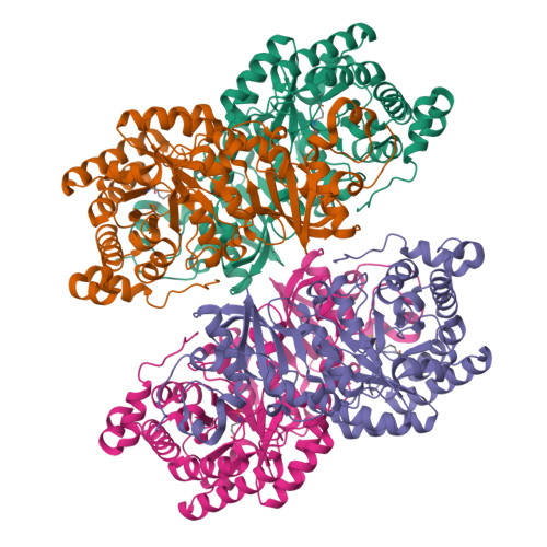
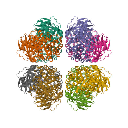
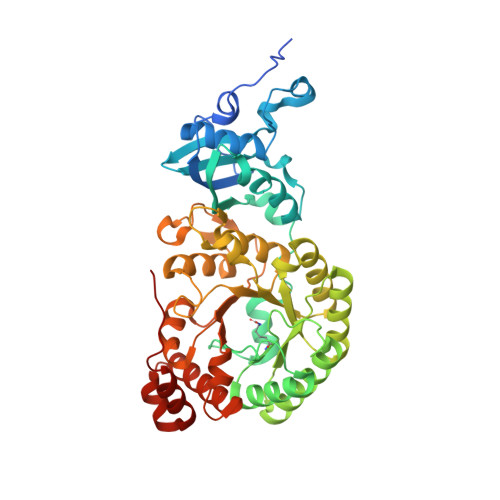
6URA

Crystal structure of RUBISCO from Promineofilum breve


Experimental Data Snapshot

  • Method: X-RAY DIFFRACTION
  • Resolution: 2.17 Å
  • R-Value Free: 0.225 
  • R-Value Work: 0.188 
  • R-Value Observed: 0.190 

Starting Model: experimental
View more details

wwPDB Validation   3D Report Full Report


Ligand Structure Quality Assessment 


This is version 1.4 of the entry. See complete history


Literature

Novel bacterial clade reveals origin of form I Rubisco.

Banda, D.M.Pereira, J.H.Liu, A.K.Orr, D.J.Hammel, M.He, C.Parry, M.A.J.Carmo-Silva, E.Adams, P.D.Banfield, J.F.Shih, P.M.

(2020) Nat Plants 6: 1158-1166

  • DOI: https://doi.org/10.1038/s41477-020-00762-4
  • Primary Citation of Related Structures:  
    6URA

  • PubMed Abstract: 

    Rubisco sustains the biosphere through the fixation of CO 2 into biomass. In plants and cyanobacteria, form I Rubisco is structurally comprised of large and small subunits, whereas all other Rubisco forms lack small subunits. The rise of the form I complex through the innovation of small subunits represents a key, yet poorly understood, transition in Rubisco's evolution. Through metagenomic analyses, we discovered a previously uncharacterized clade sister to form I Rubisco that evolved without small subunits. This clade diverged before the evolution of cyanobacteria and the origin of the small subunit; thus, it provides a unique reference point to advance our understanding of form I Rubisco evolution. Structural and kinetic data presented here reveal how a proto-form I Rubisco assembled and functioned without the structural stability imparted from small subunits. Our findings provide insight into a key evolutionary transition of the most abundant enzyme on Earth and the predominant entry point for nearly all global organic carbon.


  • Organizational Affiliation

    Department of Plant Biology, University of California, Davis, Davis, CA, USA.


Macromolecules
Find similar proteins by:  (by identity cutoff)  |  3D Structure
Entity ID: 1
MoleculeChains Sequence LengthOrganismDetailsImage
Ribulose bisphosphate carboxylase large chain
A, B, C, D
463Candidatus Promineifilum breveMutation(s): 0 
Gene Names: cbbLCFX0092_B0544
EC: 4.1.1.39
UniProt
Find proteins for A0A160T9D2 (Candidatus Promineifilum breve)
Explore A0A160T9D2 
Go to UniProtKB:  A0A160T9D2
Entity Groups  
Sequence Clusters30% Identity50% Identity70% Identity90% Identity95% Identity100% Identity
UniProt GroupA0A160T9D2
Sequence Annotations
Expand
  • Reference Sequence
Small Molecules
Ligands 2 Unique
IDChains Name / Formula / InChI Key2D Diagram3D Interactions
CAP
Query on CAP

Download Ideal Coordinates CCD File 
E [auth A],
G [auth B],
I [auth C],
K [auth D]
2-CARBOXYARABINITOL-1,5-DIPHOSPHATE
C6 H14 O13 P2
ITHCSGCUQDMYAI-ZMIZWQJLSA-N
MG
Query on MG

Download Ideal Coordinates CCD File 
F [auth A],
H [auth B],
J [auth C],
L [auth D]
MAGNESIUM ION
Mg
JLVVSXFLKOJNIY-UHFFFAOYSA-N
Modified Residues  1 Unique
IDChains TypeFormula2D DiagramParent
KCX
Query on KCX
A, B, C, D
L-PEPTIDE LINKINGC7 H14 N2 O4LYS
Experimental Data & Validation

Experimental Data

  • Method: X-RAY DIFFRACTION
  • Resolution: 2.17 Å
  • R-Value Free: 0.225 
  • R-Value Work: 0.188 
  • R-Value Observed: 0.190 
  • Space Group: C 1 2 1
Unit Cell:
Length ( Å )Angle ( ˚ )
a = 163.06α = 90
b = 162.74β = 109.57
c = 90γ = 90
Software Package:
Software NamePurpose
PHENIXrefinement
XDSdata reduction
SCALAdata scaling
PHENIXphasing

Structure Validation

View Full Validation Report


Ligand Structure Quality Assessment 


Entry History 

Deposition Data

Revision History  (Full details and data files)

  • Version 1.0: 2020-08-19
    Type: Initial release
  • Version 1.1: 2020-09-16
    Changes: Database references
  • Version 1.2: 2020-09-23
    Changes: Database references
  • Version 1.3: 2023-10-11
    Changes: Data collection, Database references, Refinement description
  • Version 1.4: 2023-11-15
    Changes: Data collection