7A8M

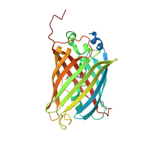
rsGreen0.7-K206A-N205G in the green-on state


Experimental Data Snapshot

  • Method: X-RAY DIFFRACTION
  • Resolution: 1.60 Å
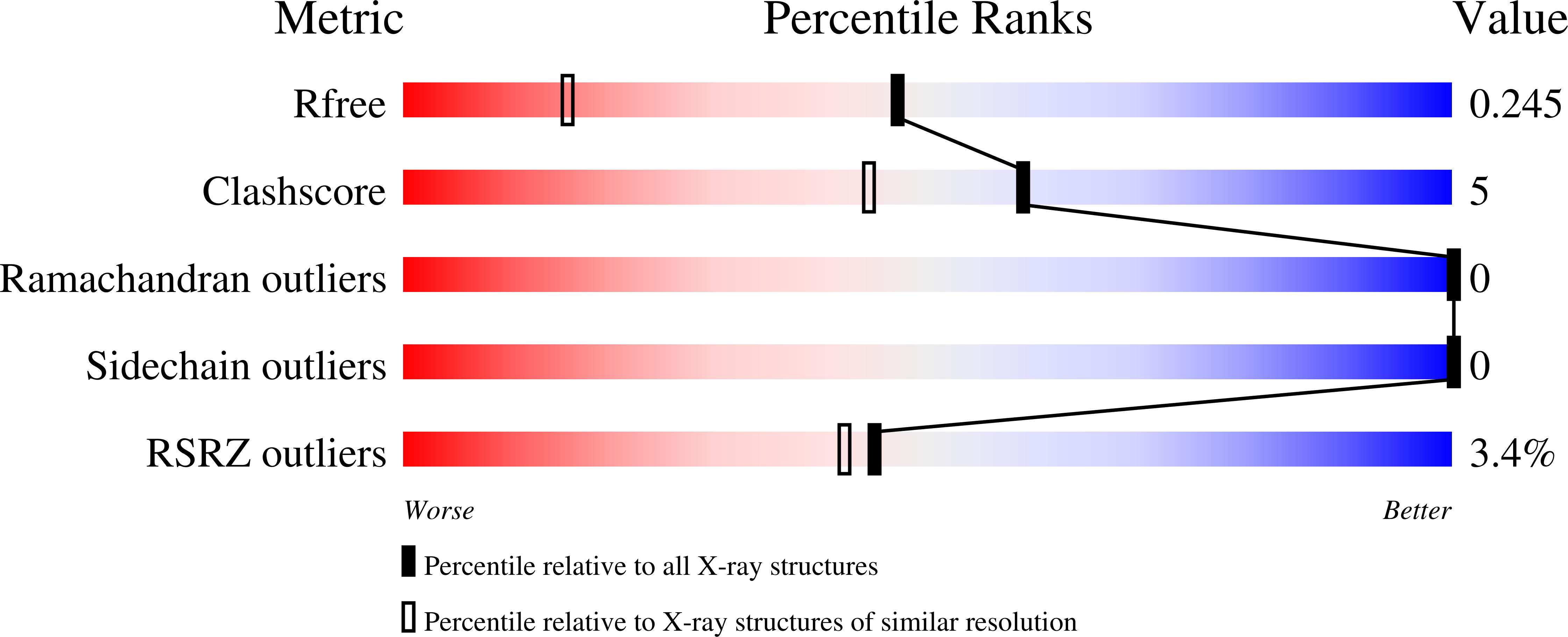
  • R-Value Free: 0.245 
  • R-Value Work: 0.202 
  • R-Value Observed: 0.204 

wwPDB Validation   3D Report Full Report


This is version 1.2 of the entry. See complete history


Literature

Structure-Function Dataset Reveals Environment Effects within a Fluorescent Protein Model System*.

De Zitter, E.Hugelier, S.Duwe, S.Vandenberg, W.Tebo, A.G.Van Meervelt, L.Dedecker, P.

(2021) Angew Chem Int Ed Engl 60: 10073-10081

  • DOI: https://doi.org/10.1002/anie.202015201
  • Primary Citation of Related Structures:  
    7A7K, 7A7L, 7A7M, 7A7N, 7A7O, 7A7P, 7A7Q, 7A7R, 7A7S, 7A7T, 7A7U, 7A7V, 7A7W, 7A7X, 7A7Y, 7A7Z, 7A80, 7A81, 7A82, 7A83, 7A84, 7A85, 7A86, 7A87, 7A88, 7A89, 7A8A, 7A8B, 7A8C, 7A8D, 7A8E, 7A8F, 7A8G, 7A8H, 7A8I, 7A8J, 7A8K, 7A8L, 7A8M, 7A8N, 7A8O

  • PubMed Abstract: 

    Anisotropic environments can drastically alter the spectroscopy and photochemistry of molecules, leading to complex structure-function relationships. We examined this using fluorescent proteins as easy-to-modify model systems. Starting from a single scaffold, we have developed a range of 27 photochromic fluorescent proteins that cover a broad range of spectroscopic properties, including the determination of 43 crystal structures. Correlation and principal component analysis confirmed the complex relationship between structure and spectroscopy, but also allowed us to identify consistent trends and to relate these to the spatial organization. We find that changes in spectroscopic properties can come about through multiple underlying mechanisms, of which polarity, hydrogen bonding and presence of water molecules are key modulators. We anticipate that our findings and rich structure/spectroscopy dataset can open opportunities for the development and evaluation of new and existing protein engineering methods.


  • Organizational Affiliation

    Department of Chemistry, KU Leuven, Celestijnenlaan 200G - box 2403, 3001, Leuven, Belgium.


Macromolecules
Find similar proteins by:  (by identity cutoff)  |  3D Structure
Entity ID: 1
MoleculeChains Sequence LengthOrganismDetailsImage
Green fluorescent protein270Aequorea victoriaMutation(s): 2 
Gene Names: gfp
UniProt
Find proteins for A0A059PIQ0 (Aequorea victoria)
Explore A0A059PIQ0 
Go to UniProtKB:  A0A059PIQ0
Entity Groups  
Sequence Clusters30% Identity50% Identity70% Identity90% Identity95% Identity100% Identity
UniProt GroupA0A059PIQ0
Sequence Annotations
Expand
  • Reference Sequence
Small Molecules
Modified Residues  1 Unique
IDChains TypeFormula2D DiagramParent
CRO
Query on CRO
A
L-PEPTIDE LINKINGC15 H17 N3 O5THR, TYR, GLY
Experimental Data & Validation

Experimental Data

  • Method: X-RAY DIFFRACTION
  • Resolution: 1.60 Å
  • R-Value Free: 0.245 
  • R-Value Work: 0.202 
  • R-Value Observed: 0.204 
  • Space Group: H 3 2
Unit Cell:
Length ( Å )Angle ( ˚ )
a = 140.869α = 90
b = 140.869β = 90
c = 73.224γ = 120
Software Package:
Software NamePurpose
XDSdata reduction
Aimlessdata scaling
PHENIXrefinement
PDB_EXTRACTdata extraction
PHENIXphasing

Structure Validation

View Full Validation Report


Entry History & Funding Information

Deposition Data


Funding OrganizationLocationGrant Number
Research Foundation - FlandersBelgium--

Revision History  (Full details and data files)

  • Version 1.0: 2021-02-17
    Type: Initial release
  • Version 1.1: 2021-05-05
    Changes: Database references
  • Version 1.2: 2024-11-13
    Changes: Data collection, Database references, Structure summary